. 2024 Nov;43(22):5421-5444.
doi: 10.1038/s44318-024-00262-7. Epub 2024 Oct 7.

Alternative splicing of PBRM1 mediates resistance to PD-1 blockade therapy in renal cancer

Affiliations

Alternative splicing of PBRM1 mediates resistance to PD-1 blockade therapy in renal cancer

Namjoon Cho et al. EMBO J. 2024 Nov.

Abstract

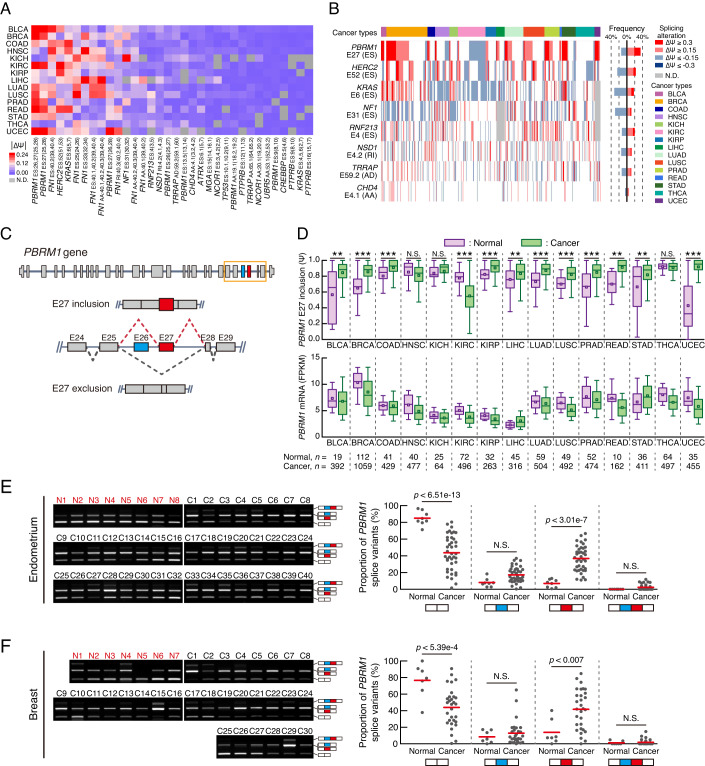
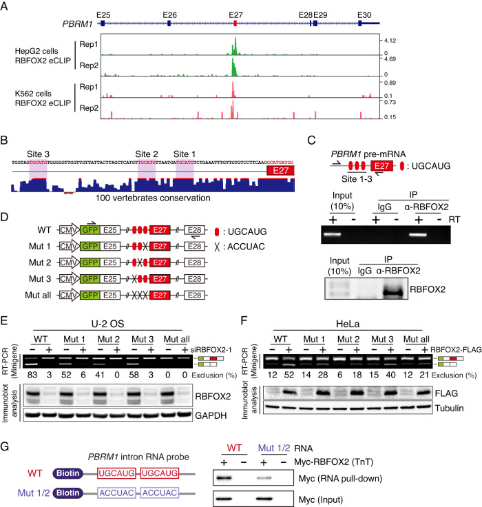
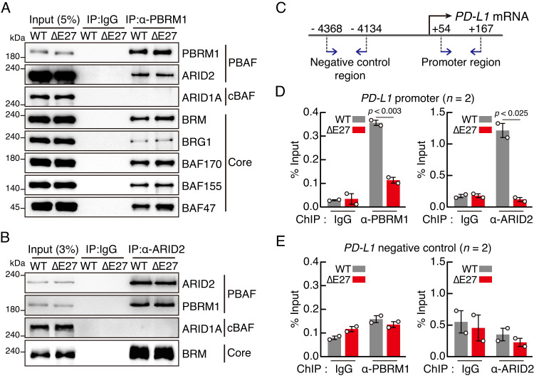
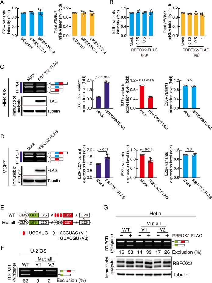
Alternative pre-mRNA splicing (AS) is a biological process that results in proteomic diversity. However, implications of AS alterations in cancer remain poorly understood. Herein, we performed a comprehensive AS analysis in cancer driver gene transcripts across fifteen cancer types and found global alterations in inclusion rates of the PBAF SWI/SNF chromatin remodeling complex subunit Polybromo 1 (PBRM1) exon 27 (E27) in most types of cancer tissues compared with those in normal tissues. Further analysis confirmed that PBRM1 E27 is excluded by the direct binding of RBFOX2 to intronic UGCAUG elements. In addition, the E27-included PBRM1 isoform upregulated PD-L1 expression via enhanced PBAF complex recruitment to the PD-L1 promoter. PBRM1 wild-type patients with clear cell renal cell carcinoma were resistant to PD-1 blockade therapy when they expressed low RBFOX2 mRNA levels. Overall, our study suggests targeting of RBFOX2-mediated AS of PBRM1 as a potential therapeutic strategy for immune checkpoint blockade.

Keywords: Alternative Splicing; Immune Checkpoint Inhibitor; PBRM1; PD-L1; RBFOX2.


Conflict of interest statement

Disclosure and competing interests statement The authors declare no competing interests.

Figures

Figure 1
Figure 1. A comprehensive pan-cancer AS analysis reveals extensive changes in the AS pattern of PBRM1 E27 in cancer tissues.
(A) Heatmap showing the AS events of cancer driver genes across 15 cancer types. The colour indicates the change in mean PSI value in cancer tissues compared with that in normal tissues. The bottom of the heatmap shows the gene name, AS type, splicing event exon, upstream exon, and downstream exon. Only the events with quantified PSI values in ≥80% of the 15 cancer types are represented in the heatmap. Ψ, PSI; ES, exon skipping; AA, alternative acceptor; AD, alternative donor; RI, retained intron; N.D., not detected. (B) Frequency of splicing alteration for selected genes from (A). The coloured line in the heatmap (left) indicates the patient with differentially spliced events between cancer tissues and adjacent normal tissues. The bar graph (right) shows the proportion of splicing alternations in each AS event. (C) Schematic representation of the AS of human PBRM1. (D) The AS patterns of PBRM1 E27 (top) or the mRNA levels of PBRM1 (bottom) in TCGA. Boxes represent the median, quartiles, 10th percentile, and 90th percentile. n, number of samples. p-values were calculated using the two-tailed Student’s t-test. (BLCA, p < 0.002; BRCA, p < 7.12e−26; COAD, p < 0.001; KIRC, p < 1.63e−33; KIRP, p < 1.62e−5; LIHC, p < 0.006; LUAD, p < 1.78e−10; LUSC, p < 1.76e−9; PRAD, p < 1.16e−8; READ, p < 0.002; STAD, p < 0.009; UCEC, p < 4.57e−11). (E, F) RT-PCR analyses of PBRM1 AS in human endometrium (E) and breast (F) samples including cancer (C) and healthy (N) tissues. The PBRM1 splice variant proportions in each tissue is presented in the dot plots (right), with red lines indicating the mean. Schematics at the bottom of the graphs represent PBRM1 splice variants. White boxes, E25 and E28; Blue box, E26; Red box, E27. p-values were calculated using one-way ANOVA with Bonferroni’s multiple comparison test. N.S., not significant. Source data are available online for this figure.
Figure 2
Figure 2. RBFOX2 regulates the E27 AS of PBRM1.
(A) Dot plots displaying splicing factors according to the correlation between mRNA levels of splicing factors and PSI values of PBRM1 E27 in cancer tissues. (B) Heatmaps of individual UCEC and BLCA samples showing an inverse correlation between the PSI values of PBRM1 E27 and mRNA levels of RBFOX2. Pearson correlation coefficient and p-value are presented. n, number of samples; Ψ, PSI. (C) Dot plot of the changes in PBRM1 E27 AS versus changes in RBFOX2 expression in different cancer types compared to those in normal tissues. Pearson correlation coefficient and p-value are presented. (D) Schematic representation of PBRM1 splice variants and its RT-PCR products. (E) Immunoblot analysis of RBFOX2 in U-2 OS and HeLa cells. (F, G) RT-PCR (top) and immunoblot (bottom) analyses showing the expression of PBRM1 splice variants in RBFOX2-knockdown U-2 OS cells (F) or in RBFOX2-FLAG-overexpressed HeLa cells (G). Coloured lines on the left of the RT-PCR results indicate the RT-PCR products, as explained in (D). Quantification of RT-PCR products for E27+ variants and E26− E27− variant are shown in bar graphs (right) (n = 3, biological replicates). Bars indicate mean ± SD. p-values were calculated using one-way ANOVA with Dunnett’s multiple comparison test. Source data are available online for this figure.
Figure 3
Figure 3. RBFOX2 directly binds to the UGCAUG elements in PBRM1 pre-mRNA.
(A) Genome browser view displaying the RBFOX2 eCLIP peaks on the PBRM1 transcripts. (B) Conserved UGCAUG elements in the upstream intronic region of PBRM1 E27 across 100 vertebrates. (C) RIP assay showing the interaction of RBFOX2 with the PBRM1 pre-mRNA. Schematic (top) representation of the primer-binding site on the PBRM1 pre-mRNA for RIP-PCR. RT-PCR (middle) and immunoblot (bottom) analyses were performed following the RIP assay. (D) Schematic representation of PBRM1 minigene constructs. (E, F) RT-PCR (top) and immunoblot (bottom) analyses for the AS pattern of the PBRM1 minigene transcripts by RBFOX2 knockdown in U-2 OS cells (E) or by RBFOX2-FLAG overexpression in HeLa cells (F). (G) RNA pull-down showing the direct binding of RBFOX2 to a biotinylated PBRM1 intron RNA probe. Source data are available online for this figure.
Figure 4
Figure 4. E27 inclusion of PBRM1 promotes PD-L1 expression.
(A) RT-PCR (top) and immunoblot (bottom) analyses showing the establishment of ΔE27 HeLa cell lines. (B) GSEA of RNA sequencing data for all gene expression in ΔE27 HeLa cells versus WT HeLa cells. Significantly enriched terms at FDR < 0.0025 and nominal p < 0.01 are represented in the bar graph. (C) GSEA of IFNγ-responsive genes in all gene transcripts of ΔE27 HeLa cells versus WT HeLa cells. The nominal p-value is presented. (D) Validation of RNA sequencing data for IFN-responsive genes by qRT-PCR analysis (n = 3, biological replicates). Bars indicate mean ± SD. p-values were calculated using two-tailed Student’s t-test. (PD-L1, p < 0.047; IFI44, p < 0.002; ISG15, p < 6.86e−5; IFIT2, p < 0.001). (E) qRT-PCR analysis of PD-L1 mRNA levels in 20 ng/ml EGF- or 40 ng/ml HGF-treated ΔE27 HeLa cells for 12 h (n = 4, biological replicates). Bars indicate mean ± SD. p-values were calculated using one-way ANOVA with Dunnett’s multiple comparison test. (Control: WT vs # 1, p < 0.004; WT vs # 2, p < 0.001; EGF: WT vs # 1, p < 1.54e−4; WT vs # 2, p < 1.40e−4; HGF: WT vs # 1, p < 2.49e−4; WT vs # 2, p < 1.88e−4). (F) qRT-PCR analysis of time-dependent PD-L1 mRNA levels in HeLa cells treated with 20 ng/ml EGF (n = 4, biological replicates). Bars indicate mean ± SD. p-values were calculated using one-way ANOVA with Dunnett’s multiple comparison test. (Control: WT vs # 1, p < 8.83e−6; WT vs # 2, p < 1.68e−5; 6 h: WT vs # 1, p < 1.84e−6; WT vs # 2, p < 9.39e−7; 12 h: WT vs # 1, p < 5.26e−8; WT vs # 2, p < 5.12e−8). (G) Immunoblot analysis of PD-L1 in 20 ng/ml EGF- and 100 ng/ml IFNγ-treated HeLa cells for 12 h following transfection with siPBRM1 for 36 h. Source data are available online for this figure.
Figure 5
Figure 5. The E27-included isoform of PBRM1 increases PBAF binding to the PD-L1 promoter region.
(A, B) IP with an anti-PBRM1 antibody (A) or an anti-ARID2 antibody (B) in WT or ΔE27 HeLa cells followed by immunoblot analysis. PBAF, polybromo-associated BAF; cBAF, canonical BAF; Core, pan-mSWI/SNF complex subunit. (C) Schematic representation of PD-L1 gene. The blue arrows indicate primer-binding sites. (D, E) ChIP-qPCR amplifying the PD-L1 promoter region (D) or negative control region (E) from an anti-PBRM1 antibody (left) or an anti-ARID2 antibody (right)-precipitated immune complexes from HeLa cell lysates (n = 2, biological replicates). Bars indicate mean ± SD. p-values were calculated using one-tailed Student’s t-test. Source data are available online for this figure.
Figure 6
Figure 6. RBFOX2-mediated AS in PBRM1 affects resistance to PD-1 blockade therapy in ccRCC.
(A) Dot plot showing PSI values of PBRM1 E27 in individual KIRC cancer tissues. n, number of samples; Ψ, PSI. (B) Heatmap showing the correlation between PSI values of PBRM1 E27 and mRNA levels of PD-L1, RBFOX2, and PBRM1 in individual KIRC tissues. n, number of samples. (C, D) Box plots showing the PD-L1 (C) or RBFOX2 (D) mRNA levels according to PSI values of PBRM1 E27 in the PBRM1 WT KIRC group or PBRM1 Mut KIRC group. Boxes represent the median, quartiles, 10th percentile, and 90th percentile. p-values were calculated using one-way ANOVA with Bonferroni’s multiple comparison test. n, number of samples; N.S., not significant. (E, F) Violin plots showing the mRNA levels of perforin (E) or granzyme B (F) according to PSI values of PBRM1 E27 in patients with KIRC. p-values were calculated using the two-tailed Student’s t-test. n, number of samples; N.S., not significant. (G) Dot plot showing individual KIRC patients with percent of samples expressing PD-L1 more than 1.75 values of normalised FPKM depending on mutation in PBRM1 (left) or VHL (right). (H, I) Kaplan–Meier survival curves of patients with ccRCC treated with nivolumab (H) or everolimus (I) from clinical trial data (Checkmate 009, 010, and 025). p-values were calculated using Log-rank test. **p < 0.002. n, number of samples; N.S., not significant. (J) Model for RBFOX2-mediated regulation of E27 AS in PBRM1 and its role in cancer immune evasion. Source data are available online for this figure.
Figure EV1
Figure EV1. RBFOX2 mRNA levels inversely correlate with PSI values of PBRM1 E27 in cancer tissues.
(A) Dot plots of splicing factors showing Pearson’s correlation coefficients between FPKM values of the splicing factors and PSI values of PBRM1 E27 in individual TCGA cancer tissues. RBFOX2 is indicated with a red dot. (B) Heatmaps showing the inverse correlation between the PSI values of PBRM1 E27 and mRNA levels of RBFOX2 in BRCA, READ, and PRAD. Pearson’s correlation coefficients and p-values are shown at the bottom of the heatmap. n, number of samples; Ψ, PSI. (C) RBFOX2 mRNA levels in endometrium cDNAs. The red lines represent the means. n, number of samples. p-values were calculated using a two-tailed Student’s t-test. Source data are available online for this figure.
Figure EV2
Figure EV2. RBFOX2 represses E27 inclusion in PBRM1 mRNA.
(A, B) Quantification of RT-PCR products shown in Fig. 2F, G for E26+ variants (left) and total PBRM1 mRNA levels in RBFOX2-knockdown U-2 OS cells (A) or in RBFOX2-FLAG-overexpressed HeLa cells (B) (n = 3, biological replicates). Bars indicate mean ± SD. (C, D) AS patterns of PBRM1 in RBFOX2 overexpressed cells. HEK293 (C) and MCF7 (D) cells were transfected with RBFOX2 expression vectors for 36 h, followed by RT-PCR (top left), immunoblot (bottom left), and qRT-PCR (right) analyses (n = 4, biological replicates). Bars indicate mean ± SD. p-values were calculated using a two-tailed Student’s t-test. N.S., not significant. (E) Schematic representation of PBRM1 minigene constructs. (F) AS pattern of PBRM1 minigene constructs in U-2 OS cells. (G) RT-PCR (top) and immunoblot (bottom) analyses for the AS pattern of PBRM1 minigene transcripts by RBFOX2-FLAG overexpression in HeLa cells. Source data are available online for this figure.
Figure EV3
Figure EV3. PBRM1 differentially affects gene expression based on E27 AS.
(A) Sanger sequencing with RT-PCR products of skipping variant, lacking E26 and E27, of PBRM1 expressed in ΔE27 HeLa cell lines. (B) Scatter plot of RNA sequencing data displaying upregulated (red) and downregulated (blue) genes in ΔE27 HeLa cells compared to those in WT HeLa cells. (C) GSEA plot of IFNα-responsive genes in all gene transcripts of ΔE27 HeLa cells versus WT HeLa cells. The nominal p-value is presented. (D) STRING protein-protein network analysis of the top 120 downregulated genes in ΔE27 HeLa cells. The lines indicate the co-expression network between proteins and the line thickness indicates the strength of data support. (E, F) qRT-PCR analysis of PD-L1 (E) or PBRM1 (F) mRNA levels. HeLa cells were treated with 20 ng/ml EGF and 100 ng/ml IFNγ for 12 h following transfection with siPBRM1 for 36 h (n = 3, biological replicates). Bars indicate mean ± SD. p-values were calculated using one-way ANOVA with Dunnett’s multiple comparison test. (G, H) Immunoblot (left) and qRT-PCR (right) analyses confirming PD-L1 expression levels following knockdown of the E27-included PBRM1 isoforms using siRNA targeting PBRM1 E27 sequences (siE27-PBRM1) in HeLa (G) or MDA-MB-231 cells (H). n = 3, biological replicates. Bars indicate mean ± SD. p-values were calculated using two-tailed Student’s t-test. N.S., not significant. (I) PD-L1 promoter reporter assay in WT and ΔE27 HeLa cells. The schematic shows the PD-L1 promoter reporter vector (top), and the graph presents the results of firefly luciferase (Fluc) activities normalised to renilla luciferase (Rluc) activities (bottom). n = 3, biological replicates. Lines indicate the mean. p-values were calculated using one-way ANOVA with Bonferroni’s multiple comparison test. ISRE, Interferon-sensitive response element. (J) Immunofluorescence analysis of PBRM1 (green) and DAPI (nuclei; blue) in WT and ΔE27 HeLa cells. Scale bars, 20 μm. Source data are available online for this figure.
Figure EV4
Figure EV4. SSO-induced E27 exclusion of PBRM1 suppresses cancer immune evasion.
(A) NK-92 cell cytotoxicity against ΔE27 HeLa cells. HeLa cells were co-cultured with NK-92 cells for 7 h (n = 3, biological replicates). p-values were calculated using one-tailed Student’s t-test. (B) Design of PBRM1 SSO for induction of E27 exclusion in PBRM1. (C) NK-92 cell cytotoxicity against PBRM1 SSO-transfected cancer cells (top). NK-92 cells were co-cultured with cancer cells at an effector:target ratio of 20:1 for 7 h (n = 3, biological replicates). SSO-induced PBRM1 E27 exclusion was confirmed using RT-PCR (bottom). Bars indicate mean ± SD. p-values were calculated using one-tailed Student’s t-test. (D) RT-PCR analysis confirmed PBRM1 E27 exclusion by PBRM1 SSO transfection into B16-F10 cells used for subcutaneous injection into mice. (E, F) Tumour volume (E) and representative images of tumour (F) from C57BL/6 mice injected subcutaneously with control SSO- (n = 9, individual mice) or PBRM1 SSO-transfected B16-F10 cells (n = 10, individual mice) on day 9. The p-value was calculated using two-tailed Student’s t-test. Scale bar, 1 cm. Source data are available online for this figure.
Figure EV5
Figure EV5. E27 inclusion rates in PBRM1 correlate with PD-L1 expression in PBRM1 WT ccRCC but not in PBRM1 Mut ccRCC.
(A) Mutation frequency in the PBRM1 gene across patients with KIRC in the PBRM1 Mut group. Boxes indicate cancer tissues, with colours representing the four somatic mutation statuses. (B) Box plot showing PBRM1 mRNA level in normal and ccRCC tissues. Boxes represent the median, quartiles, 10th percentile, and 90th percentile. n, number of samples. p-values were calculated by the one-way ANOVA with Bonferroni’s multiple comparison test. (C, D) Scatter plot of PBRM1 E27 PSI versus PD-L1 mRNA level (C) or RBFOX2 mRNA level (D) in individual PBRM1 WT ccRCC (left) or PBRM1 Mut ccRCC (right) cancer tissues. Pearson correlation coefficients and p-values are presented. n, number of samples; Ψ, PSI. (E, F) Kaplan–Meier survival curves of patients with ccRCC treated with nivolumab (E) or everolimus (F). Patients with ccRCC possessing the non-mutant VHL gene and a low VHL mRNA level (below 12%) were not classified in the VHL WT group. n, number of samples. (G) Pie charts showing the ratios of ccRCC patients with high and low PD-L1 levels in high (RBFOX2 low and PBRM1 WT) or low (all others) E27 functional groups. Patients with ccRCC treated with nivolumab or everolimus were classified. n, number of samples. The p-value was calculated using chi-squared test. (H) Kaplan–Meier survival curve of ccRCC patients treated with nivolumab, stratified by PD-L1 expression status. n, number of samples. N.S., not significant.

Similar articles

Cited by

References

    1. Bailey MH, Tokheim C, Porta-Pardo E, Sengupta S, Bertrand D, Weerasinghe A, Colaprico A, Wendl MC, Kim J, Reardon B et al (2018) Comprehensive characterization of cancer driver genes and mutations. Cell 173:371–385 - PMC - PubMed
    1. Barbosa-Morais NL, Irimia M, Pan Q, Xiong HY, Gueroussov S, Lee LJ, Slobodeniuc V, Kutter C, Watt S, Colak R et al (2012) The evolutionary landscape of alternative splicing in vertebrate species. Science 338:1587–1593 - PubMed
    1. Bludau I, Aebersold R (2020) Proteomic and interactomic insights into the molecular basis of cell functional diversity. Nat Rev Mol Cell Bio 21:327–340 - PubMed
    1. Bolger AM, Lohse M, Usadel B (2014) Trimmomatic: a flexible trimmer for Illumina sequence data. Bioinformatics 30:2114–2120 - PMC - PubMed
    1. Braun DA, Hou Y, Bakouny Z, Ficial M, Sant’ Angelo MS, Forman J, Ross-Macdonald P, Berger AC, Jegede OA, Elagina L et al (2020) Interplay of somatic alterations and immune infiltration modulates response to PD-1 blockade in advanced clear cell renal cell carcinoma. Nat Med 26:909–918 - PMC - PubMed

MeSH terms