

Neuroimaging studies of resting-state functional magnetic resonance imaging in eating disorders
- PMID: 39375605
- PMCID: PMC11460144
- DOI: 10.1186/s12880-024-01432-z
Neuroimaging studies of resting-state functional magnetic resonance imaging in eating disorders
Abstract
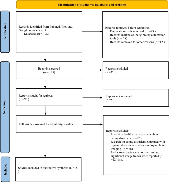
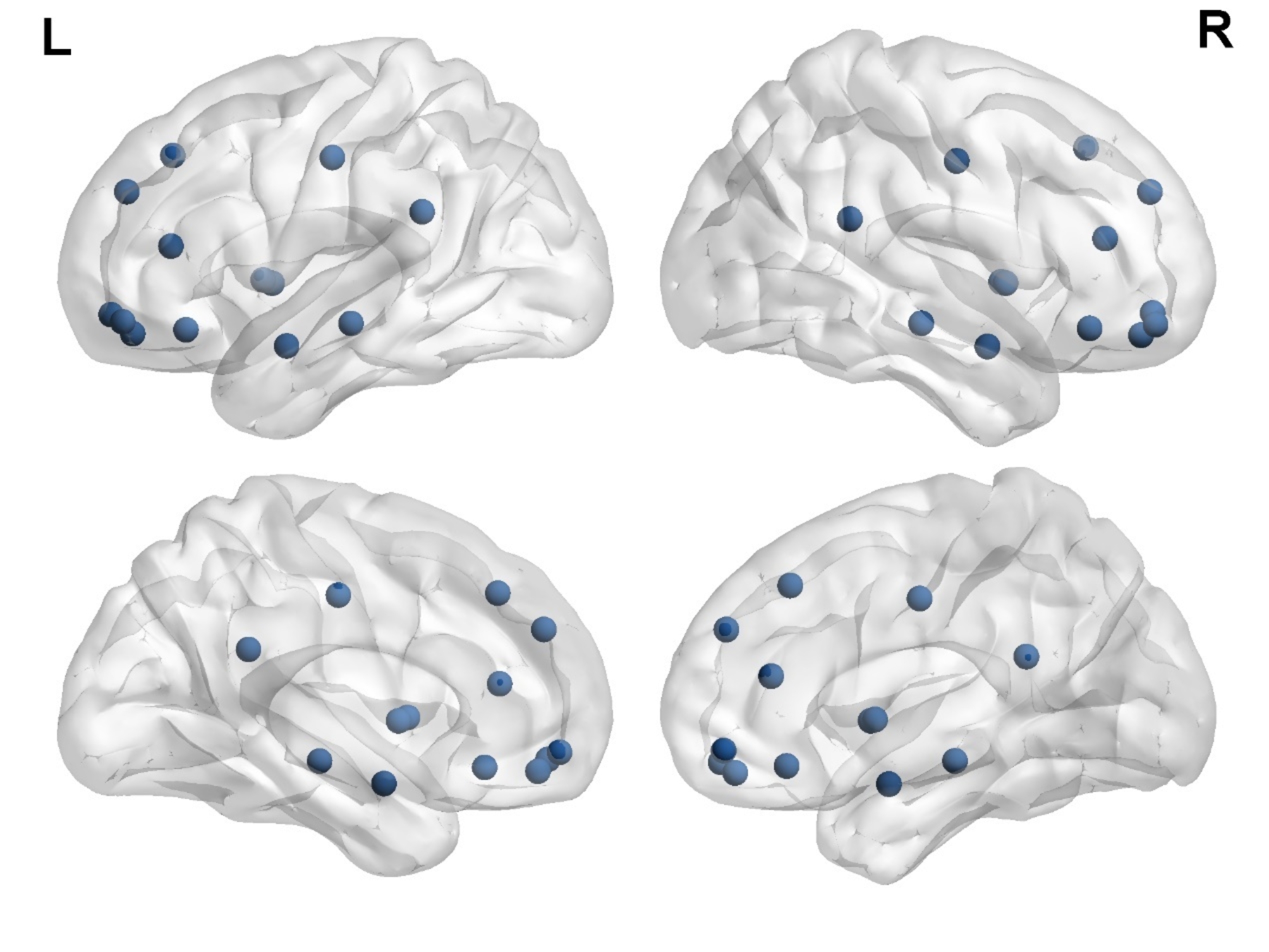
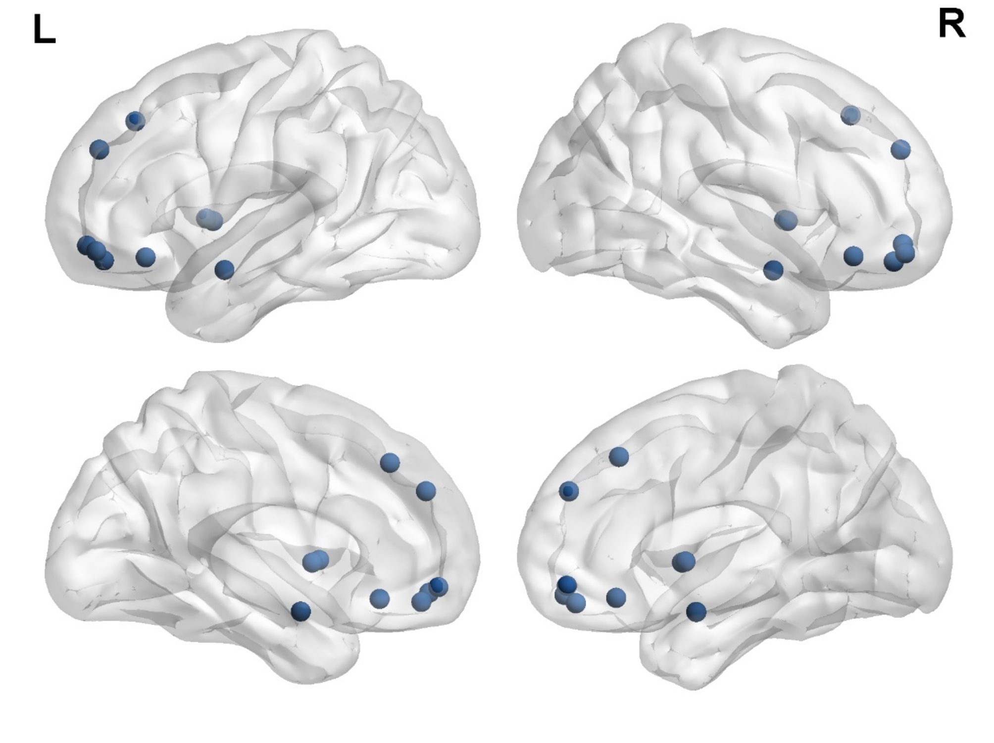
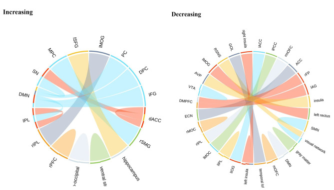
Eating disorders (EDs), including anorexia nervosa (AN), bulimia nervosa (BN), binge-eating disorder (BED), and pica, are psychobehavioral conditions characterized by abnormal eating behaviors and an excessive preoccupation with weight and body shape. This review examines changes in brain regions and functional connectivity in ED patients over the past decade (2013-2023) using resting-state functional magnetic resonance imaging (rs-fMRI). Key findings highlight alterations in brain networks such as the default mode network (DMN), central executive network (CEN), and emotion regulation network (ERN). In individuals with AN, there is reduced functional connectivity in areas associated with facial information processing and social cognition, alongside increased connectivity in regions linked to sensory stimulation, aesthetic judgment, and social anxiety. Conversely, BED patients show diminished connectivity in the dorsal anterior cingulate cortex within the salience network and increased connectivity in the posterior cingulate cortex and medial prefrontal cortex within the DMN. These findings suggest that rs-fMRI could serve as a valuable biomarker for assessing brain function and predicting treatment outcomes in EDs, paving the way for personalized therapeutic strategies.
Keywords: Brain networks; Eating disorders; Functional connectivity; Neuroimaging; Resting-state functional magnetic resonance imaging.
© 2024. The Author(s).
Conflict of interest statement
The authors declare no competing interests.
Figures

Similar articles
-
Neural Network Alterations Across Eating Disorders: A Narrative Review of fMRI Studies.Curr Neuropharmacol. 2018;16(8):1150-1163. doi: 10.2174/1570159X15666171017111532. Curr Neuropharmacol. 2018. PMID: 29046154 Free PMC article. Review.
-
Altered functional connectivity in binge eating disorder and bulimia nervosa: A resting-state fMRI study.Brain Behav. 2019 Feb;9(2):e01207. doi: 10.1002/brb3.1207. Epub 2019 Jan 15. Brain Behav. 2019. PMID: 30644179 Free PMC article.
-
Neuroimaging and neuromodulation approaches to study eating behavior and prevent and treat eating disorders and obesity.Neuroimage Clin. 2015 Mar 24;8:1-31. doi: 10.1016/j.nicl.2015.03.016. eCollection 2015. Neuroimage Clin. 2015. PMID: 26110109 Free PMC article. Review.
-
Effects of integrated hospital treatment on the default mode, salience, and frontal-parietal networks in anorexia nervosa: A longitudinal resting-state functional magnetic resonance imaging study.PLoS One. 2023 May 30;18(5):e0283318. doi: 10.1371/journal.pone.0283318. eCollection 2023. PLoS One. 2023. PMID: 37253028 Free PMC article.
-
Default mode network mechanisms of transcranial magnetic stimulation in depression.Biol Psychiatry. 2014 Oct 1;76(7):517-26. doi: 10.1016/j.biopsych.2014.01.023. Epub 2014 Feb 5. Biol Psychiatry. 2014. PMID: 24629537 Free PMC article.
Cited by
-
Dynamic cross-lagged effects between healthy lifestyles and multimorbidity among middle-aged and older adults in China.BMC Public Health. 2025 Jun 7;25(1):2132. doi: 10.1186/s12889-025-23397-6. BMC Public Health. 2025. PMID: 40483398 Free PMC article.
-
Neuroimaging and machine learning in eating disorders: a systematic review.Eat Weight Disord. 2025 Jun 1;30(1):46. doi: 10.1007/s40519-025-01757-w. Eat Weight Disord. 2025. PMID: 40450619 Free PMC article. Review.
-
Case Report: The intersection of psychiatry and medicine: diagnostic and ethical insights from case studies.Front Psychiatry. 2025 Apr 22;16:1576179. doi: 10.3389/fpsyt.2025.1576179. eCollection 2025. Front Psychiatry. 2025. PMID: 40330647 Free PMC article.
-
Considerations for informing precision psychiatry in eating disorders: Foundations for future practice.J Eat Disord. 2025 Jul 30;13(1):162. doi: 10.1186/s40337-025-01351-6. J Eat Disord. 2025. PMID: 40739669 Free PMC article.
-
Biological treatments for co-occurring eating disorders and psychological trauma: a systematic review.Front Psychiatry. 2025 Feb 21;16:1523269. doi: 10.3389/fpsyt.2025.1523269. eCollection 2025. Front Psychiatry. 2025. PMID: 40104330 Free PMC article.
References
-
- Steinglass J, Walsh BT. Habit learning and anorexia nervosa: a cognitive neuroscience hypothesis. Int J Eat Disord. 2006;39(4):267–75. - PubMed
Publication types
MeSH terms
Grants and funding
LinkOut - more resources
Full Text Sources
Medical
Miscellaneous
