. 2024 Oct 7;24(1):361.
doi: 10.1186/s12906-024-04667-z.

Exploring the complex immunomodulatory effects and gut defense via oral administration of Astragali radix water extract to normal mice

Affiliations

Exploring the complex immunomodulatory effects and gut defense via oral administration of Astragali radix water extract to normal mice

Mi-Gi Lee et al. BMC Complement Med Ther. .

Abstract

Background: Astragali radix (AR) is one of the most widely used traditional Chinese herbal medicines. It exhibits diverse biological activities, including immunomodulatory and anti-inflammatory properties; however, some of its activities have only been demonstrated in vitro.

Objective: To examine the effects of orally administered AR extract on immune cells and the intestine under physiological conditions, which bridges the gap between previously observed in vitro outcomes and in vivo results.

Methods: AR extract was prepared by hot water extraction. Three separate animal experiments were conducted to isolate macrophages, splenocytes, and the small intestine epithelium. For the macrophage preparation experiment, an intraperitoneal injection of sterile thioglycolate was administered. The mice received oral AR extract at doses of 0.1, 0.5, or 2.5 g/kg for ten days. At the end of each experiment, cells or tissues were isolated. A portion of macrophages and splenocytes were analyzed for the phenotypic changes. The remaining cells were cultured and stimulated with lipopolysaccharide (LPS) or mitogen ex vivo to assess activation status, proliferation, and cytokine production. Samples of the intestine were subjected to real-time RT-PCR.

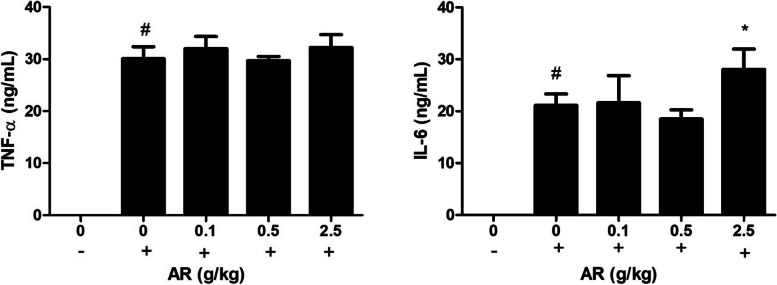
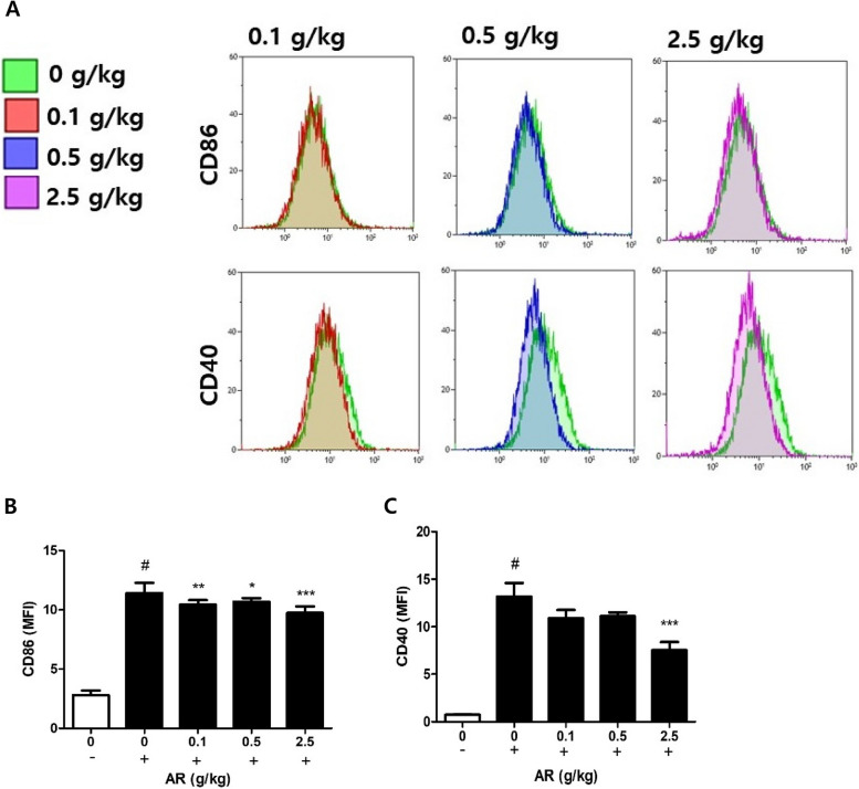
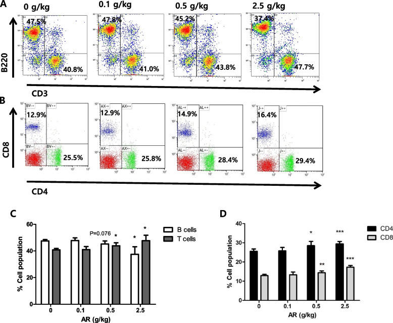
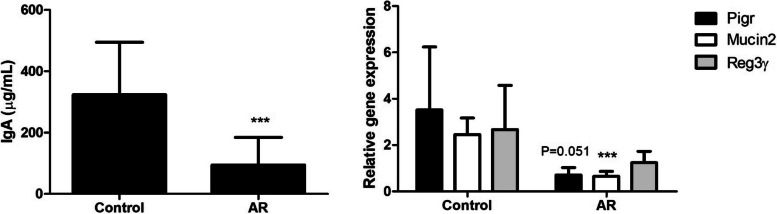
Results: Peritoneal macrophages from AR-treated mice exhibited increased expression of scavenger receptors, including SRA and CD36. Stimulation of these macrophages ex vivo with LPS selectively modulated the inflammatory response, including reduced expression of the costimulatory molecules CD40 and CD86, which are important for T cell responses, without affecting TNF-α and IL-6 production. Splenocytes from AR-treated mice exhibited a dose-dependent increase in CD4 and CD8 T cells; however, stimulation with mitogen decreased T cell proliferation and reduced IFN-γ production, which is essential for macrophage activation. An analysis of the small intestinal epithelium revealed an attenuated antimicrobial response, including reduced IgA content in the lumen and decreased expression of mucin-2 and polymeric Ig receptor genes.

Conclusion: The response of immune cells following oral treatment with AR extract did not replicate the previously documented in vitro findings. Immune cells and intestinal epithelium from mice administered oral AR extract exhibited a selective anti-inflammatory phenotype. The overall findings indicate that the systemic effects after oral administration of AR extract include reduced sensitivity to inflammatory insults.

Keywords: Antimicrobial; Astragali; Intestine; Macrophages; Splenocytes; Th1 response.


Conflict of interest statement

The authors declare no competing interests.

Figures

Fig. 1
Fig. 1
Enhanced scavenger receptor expression after the oral administration of Astragali radix extract (AR). Mice were orally administered AR (0.1, 0.5, or 2 g/kg) for 10 days. A, B Peritoneal macrophages were isolated from thioglycolate-injected mice and double-stained with FITC-conjugated anti-CD11b antibody (ab) and PE-conjugated anti-CD36 ab or FITC-conjugated anti-SRA ab and PE-conjugated anti-CD11b ab. Representative dot plots are shown. C, D Bars represent the percentage of the double-stained cell populations and the mean fluorescent intensity (MFI) of CD36 and SRA (n = 6 per group). * P < 0.05, *** P < 0.005 versus Control (0 g/kg)
Fig. 2
Fig. 2
The oral administration of AR did not suppress the production of LPS-induced inflammatory cytokines in macrophages. Macrophages isolated from the control and AR groups were stimulated with LPS (100 ng/mL) for 24 h. Tumor necrosis factor (TNF)-α and interleukin (IL)-6 levels in the supernatant were measured by ELISA (n = 6 per group). # P < 0.005 versus Control (0 g/kg, –), * P < 0.05 versus Control (0 g/kg, +)
Fig. 3
Fig. 3
The oral administration of AR decreased the surface expression of LPS-induced costimulatory molecules. Macrophages isolated from the control and AR groups were stimulated with 100 ng/mL LPS for 24 h and stained with PE-conjugated anti-CD86 ab or FITC-conjugated anti-CD40 ab. A, B Representative overlaid histograms are shown. C Bars represent the percentage of the MFI of CD86 and CD40 (n = 6 per group). (–): without LPS, ( +): LPS treatment. # P < 0.005 versus Control (–), * P < 0.05, ** P < 0.01, *** P < 0.005 versus Control ( +)
Fig. 4
Fig. 4
Dose-dependent increase in splenic T cells after the oral administration of AR. Splenocytes were isolated from the control and AR groups and double-stained with FITC-conjugated anti-CD3 antibody and PE-conjugated anti-B220 antibody for T cell and B cell markers, respectively, or FITC-conjugated anti-CD4 antibody and PE-conjugated anti-CD8 antibody. A, B Representative dot plots are shown. C, D Bars represent the percentage of CD4( +) cells or CD8( +) cells (n = 5 per group). * P < 0.05, P < 0.01, *** P < 0.005 versus Control
Fig. 5
Fig. 5
Alterations in anti-CD3 ab-induced proliferation and cytokine production in splenocytes after the oral administration of AR. Splenocytes isolated from control and AR groups were stimulated with anti-CD3 antibody (2 µg/mL) for 48 h (n = 5 per group). A The proliferation of the splenocytes was measured using the MTS assay. BD Cytokine production at 48 h of stimulation was measured by ELISA. (–): without anti-CD3 ab, ( +): anti-CD3 ab treatment. # P < 0.01, ## P < 0.001 versus Control (–), * P < 0.05, ** P < 0.01, *** P < 0.005 versus Control ( +)
Fig. 6
Fig. 6
Reduced IgA and antimicrobial gene expression in the small intestine of mice treated with AR. A Luminal fluid was obtained from the small intestine, and IgA was analyzed by ELISA (n = 10 per group). B Epithelial cells were isolated from the small intestine. RNA was extracted, and qPCR was performed. Target genes were normalized to GAPDH levels (n = 5 per group). *** P < 0.005 versus Control

Similar articles

Cited by

References

    1. Fitzgerald KA, Kagan JC. Toll-like Receptors and the Control of Immunity. Cell. 2020;180:1044–66. - PMC - PubMed
    1. Chen S, Saeed A, Liu Q, Jiang Q, Xu H, Xiao GG, et al. Macrophages in immunoregulation and therapeutics. Signal Transduct Target Ther. 2023;8:207. - PMC - PubMed
    1. Bodmer M, Itano A, McInnes I. Harnessing the small intestinal axis to resolve systemic inflammation. Front Immunol. 2022;13:1060607. - PMC - PubMed
    1. Shulzhenko N, Morgun A, Hsiao W, Battle M, Yao M, Gavrilova O, et al. Crosstalk between B lymphocytes, microbiota and the intestinal epithelium governs immunity versus metabolism in the gut. Nat Med. 2011;17:1585–93. - PMC - PubMed
    1. Gommerman JL, Rojas OL, Fritz JH. Re-thinking the functions of IgA(+) plasma cells. Gut Microbes. 2014;5:652–62. - PMC - PubMed

LinkOut - more resources