. 2024 Oct 7;22(1):911.
doi: 10.1186/s12967-024-05675-w.

FANCA promotes lung adenocarcinoma progression and is a potential target for epitope vaccine immunotherapy

Affiliations

FANCA promotes lung adenocarcinoma progression and is a potential target for epitope vaccine immunotherapy

Yanli Kang et al. J Transl Med. .

Abstract

Background: FANCA mutations have been detected in a variety of cancers and found to be pro-carcinogenic. However, no functional studies have been identified regarding the involvement of FANCA in the occurrence and the immune response of LUAD.

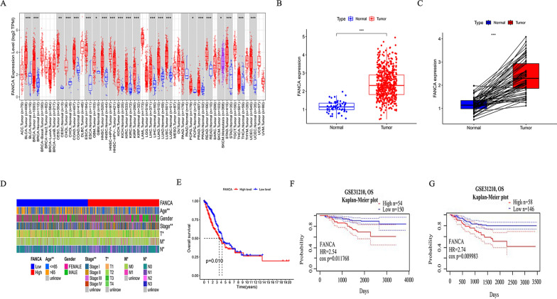
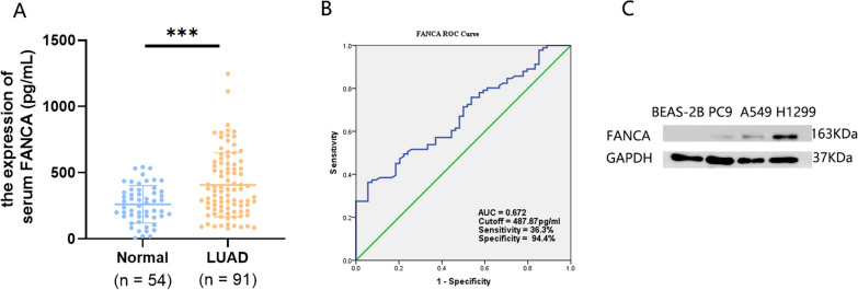
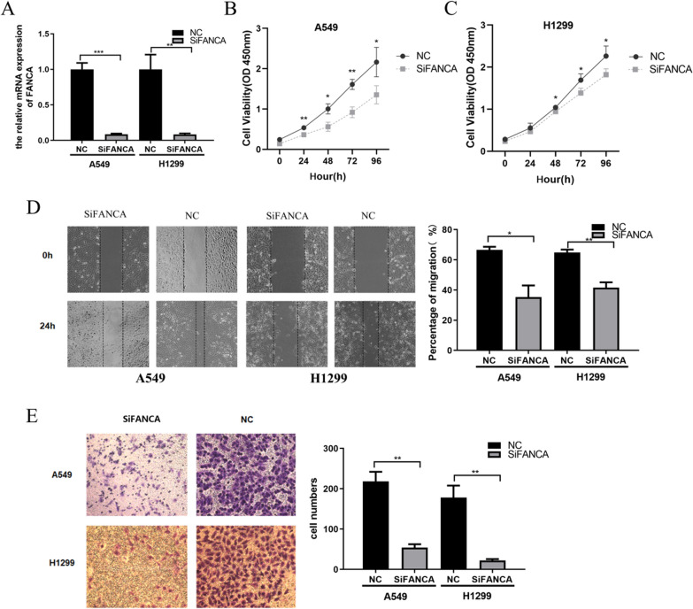
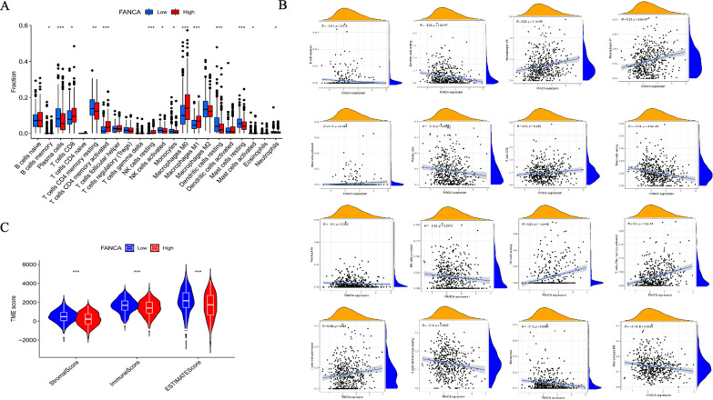
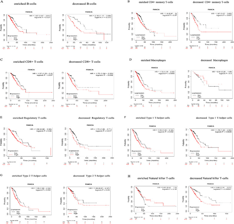
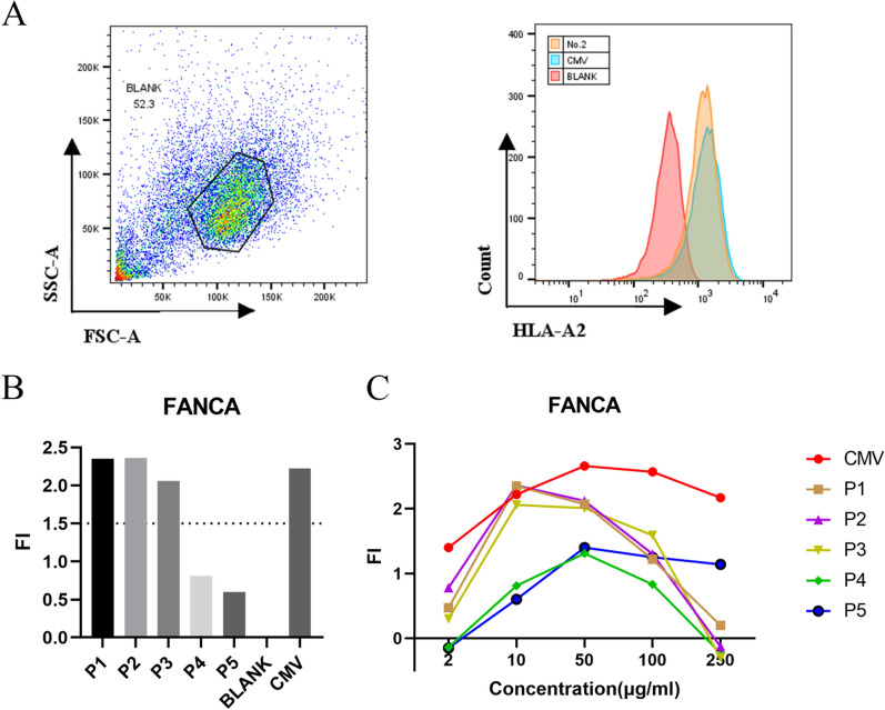
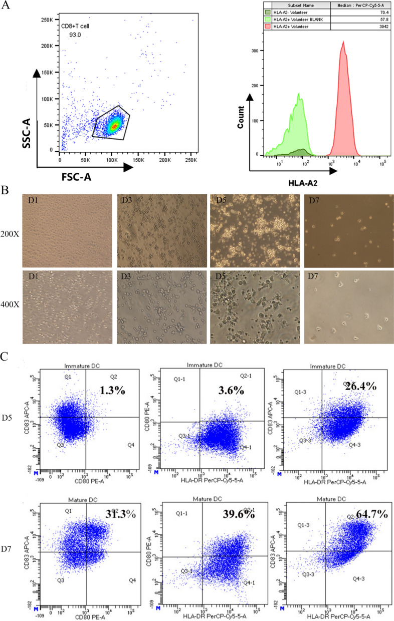
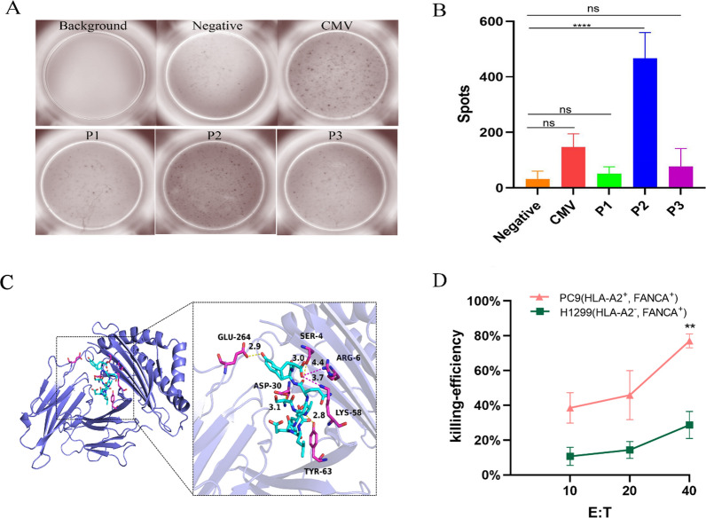
Methods: The mRNA expression and overall survival rates of FANCA were evaluated by the TIMER, PrognoScan and TCGA database in LUAD tissues, and FANCA expression was further validated by clinical serum samples using ELISA. The correlation between FANCA and immune infiltration level was investigated via TISIDB database and CIBERSORT algorithm. The Kaplan-Meier plotter was used to further evaluate the prognostic value based on the expression levels of FANCA in related immune cells. Then, the influence of FANCA knockout on the proliferation, migration, and invasion of A549 and H1299 cells was validated using CCK8, cloning formation, and Transwell assays. Subsequently, HLA-A2-restricted FANCA antigenic peptides were predicted and synthesized by NetMHC4.0 and SYFPEITHI, and DCs were induced and cultured in vitro. Finally, DCs loaded with HLA-A2-restricted FANCA antigenic peptides were co-cultured with autologous peripheral blood lymphocyte to generate specific CTLs. The killing effects of different CTLs on LUAD cells were studied.

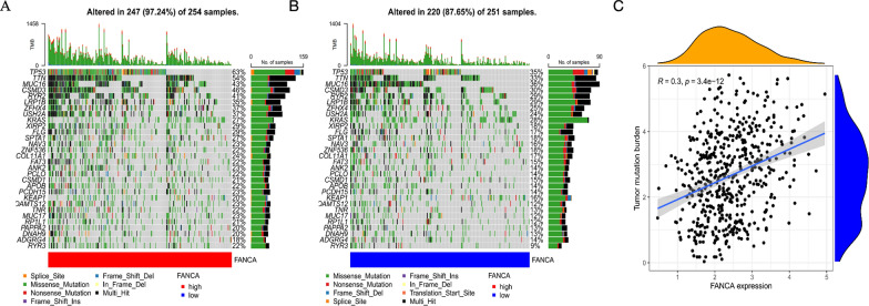
Results: The results showed that high levels of FANCA in patients with LUAD were significantly correlated with worse OS survival, which was correlated with age, clinical stage, pathological T stage, M stage, and N stage in LUAD. Knockdown of FANCA in A549 and H1299 cells significantly inhibited proliferation, metastasis, and invasion in vitro. In addition, FANCA was significantly related to immune infiltrate, genomic alterations and TMB. FANCA expression infuenced the prognosis of LUAD patients by directly affecting immune cell infltration. Finally, HLA-A2-restricted FANCA antigenic peptides were synthesized. And FANCA 146-154 (SLLEFAQYL) antigenic peptide exhibit a stronger affinity for DCs, and induce CTLs to produce stronger targeted killing ability for LUAD cells at an effector-to-target ratio of 40:1.

Conclusion: These results demonstrated that the elevation of FANCA promotes malignant phenotype of LUAD, and the potential peptide P2 (SLLEFAQYL) derived from FANCA may be used as an epitope vaccine for the treatment of LUAD.


Conflict of interest statement

The authors declared no competing interests that could potentially infuence or bias the outcomes of this research.

Figures

Fig. 1
Fig. 1
Analysis the mRNA level and prognosis of FANCA in lung adenocarcinoma. A The mRNA expression levels of FANCA in pan-cancer using TIMER database. Comparison of FANCA mRNA expression in lung adenocarcinom with adjacent normal lung tissues (B) and matched adjacent normal lung tissues C using TCGA database. D The correlation between FANCA expression and clinicopathological parameters was analyzed in TGGA database using R language. E The correlation between FANCA expression and OS was analyzed by TISIDB in LUAD. GSE31210 cohort was used to analyze the correlation between FANCA expression with OS (F) and RFS (G) in patients with LUAD by PrognoScan database (*P < 0.05, **P < 0.01, ***P < 0.001)
Fig. 2
Fig. 2
FANCA protein levels in LUAD patient serum and LUAD cells. A The protein level of FANCA in LUAD serum and healthy person’s serum. B ROC curve of serum FANCA. C The protein level of FANCA in LUAD cells
Fig. 3
Fig. 3
FANCA promotes LUAD cell proliferation, migration and invasion. A Relative expression level of FANCA after siRNA transient knockdown in A549 and H1299 cells. B-C The low expression of FANCA suppressed A549 and H1299 cells proliferation by CCK8 viability analysis. D The low expression of FANCA significantly suppressed A549 and H1299 cells migration by wound healing experiment. EThe low expression of FANCA suppressed cell invasion by transwell assay. (*P < 0.05, **P < 0.01, ***P < 0.001)
Fig. 4
Fig. 4
The relationship between FANCA expression and somatic variants and TMBscore of LUAD patients in TCGA database. The oncoPrint plots of gene mutant frequency in high (A) and low (B) FANCA expression group. Correlation between FANCA expression and Tumor Mutuation Burden (C)
Fig. 5
Fig. 5
FANCA correlates with infiltration of immune cells. A Box plots of 22 immune cell infiltration levels between the high and low FANCA groups; B Correlation between FANCA expression and the immune cell infiltration levels; C Correlation between FANCA expression and the Stromal score, immune score and ESTIMATE score in the high and low expression groups
Fig. 6
Fig. 6
Comparison of Kaplan–Meier survival curves of the high and low expression of FANCA in LUAD based on immune cell subgroups. A-H Relationships between FANCA of different immune cell subgroup and prognoses in in LUAD patients
Fig. 7
Fig. 7
Screening of FANCA peptides by the T2 binding assay. A Representative multiparameter flow cytometry scatter diagram for T2 cell affinity detection. B Fluorescence intensity (FI) value of the five peptides at a concentration of 10 µg/mL. C FI values of these peptides across a range of concentrations
Fig. 8
Fig. 8
Identification of HLA-A2 positive healthy volunteers and induction of DCs in vitro. A Expression of HLA-A2 on the surface of CD8 + T cells in peripheral blood of healthy volunteers. B Morphological of dendritic cells which cultured in vitro. C Flow cytometry was utilized to analyze the expression levels of CD80, CD83, and HLA-DR as dendritic cells progress from an immature to a mature state
Fig. 9
Fig. 9
HLA-A2-Restricted FANCA peptide P2-induced CTLs secrete IFN-γ and specifically lyse LUAD cell line PC9 in vitro. FANCA peptide-induced CTLs secrete IFN-γ in vitro. A Images result of the ELISPOT assay (B) Spot counts of the ELISPOT assay. Cytotoxic activity of FANCA peptide-induced CTLs to target cells detected by LDH release. C The interactions between FANCA protein and P2 ligand, all functional residues were identified and classified according to their interactions. D The killing efficiency of P2-induced specific CTLs against H1299 and PC9. (**P < 0.01, ****P < 0.0001)

Similar articles

Cited by

References

    1. Thai AA, Solomon BJ, Sequist LV, Gainor JF, Heist RS. Lung cancer. Lancet. 2021;398(10299):535–54. 10.1016/S0140-6736(21)00312-3. - PubMed
    1. Blandin Knight S, Crosbie PA, Balata H, Chudziak J, Hussell T, Dive C. Progress and prospects of early detection in lung cancer. Open Biol. 2017;7(9): 170070. 10.1098/rsob.170070. - PMC - PubMed
    1. Sorin M, Rezanejad M, Karimi E, Fiset B, Desharnais L, Perus LJM, Milette S, Yu MW, Maritan SM, Doré S, Pichette É, Enlow W, Gagné A, Wei Y, Orain M, Manem VSK, Rayes R, Siegel PM, Camilleri-Broët S, Fiset PO, Desmeules P, Spicer JD, Quail DF, Joubert P, Walsh LA. Single-cell spatial landscapes of the lung tumour immune microenvironment. Nature. 2023;614(7948):548–54. 10.1038/s41586-022-05672-3. - PMC - PubMed
    1. Denisenko TV, Budkevich IN, Zhivotovsky B. Cell death-based treatment of lung adenocarcinoma. Cell Death Dis. 2018;9(2):117. 10.1038/s41419-017-0063-y. - PMC - PubMed
    1. Chen D, Sha H, Hu T, Dong S, Zhang J, Liu S, Cao H, Ma R, Wu Y, Jing C, Wang Z, Wu J, Feng J. Cytokine-induced killer cells as a feasible adoptive immunotherapy for the treatment of lung cancer. Cell Death Dis. 2018;9(3):366. 10.1038/s41419-018-0404-5. - PMC - PubMed

MeSH terms

Substances