. 2024 Oct 7;22(1):478.
doi: 10.1186/s12964-024-01861-x.

Senescent cells promote breast cancer cells motility by secreting GM-CSF and bFGF that activate the JNK signaling pathway

Affiliations

Senescent cells promote breast cancer cells motility by secreting GM-CSF and bFGF that activate the JNK signaling pathway

Nan Wang et al. Cell Commun Signal. .

Abstract

Background: Cellular senescence can be induced in mammalian tissues by multiple stimuli, including aging, oncogene activation and loss of tumor suppressor genes, and various types of stresses. While senescence is a tumor suppressing mechanism when induced within premalignant or malignant tumor cells, senescent cells can promote cancer development through increased secretion of growth factors, cytokines, chemokines, extracellular matrix, and degradative enzymes, collectively known as senescence-associated secretory phenotype (SASP). Previous studies indicated that senescent cells, through SASP factors, stimulate tumor cell invasion that is a critical step in cancer cell metastasis.

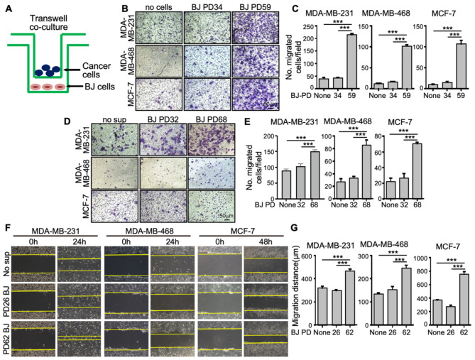
Methods: In the current study, we investigated the effect of senescent cells on the motility of breast cancer cells, which is another key step in cancer cell metastasis. We analyzed the motility of breast cancer cells co-cultured with senescent cells in vitro and metastasis of the breast cancer cells co-injected with senescent cells in orthotopic xenograft models. We also delineated the signaling pathway mediating the effect of senescent cells on cancer cell motility.

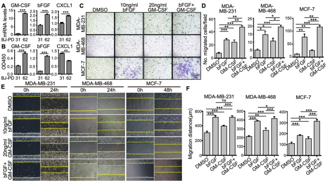
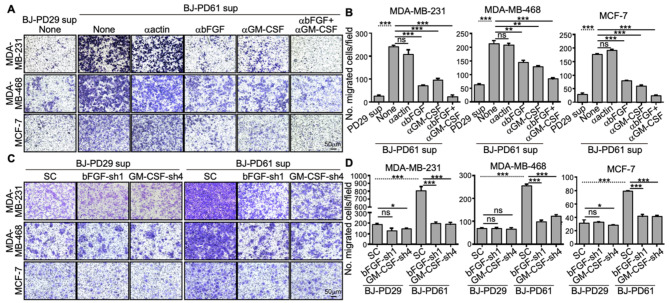
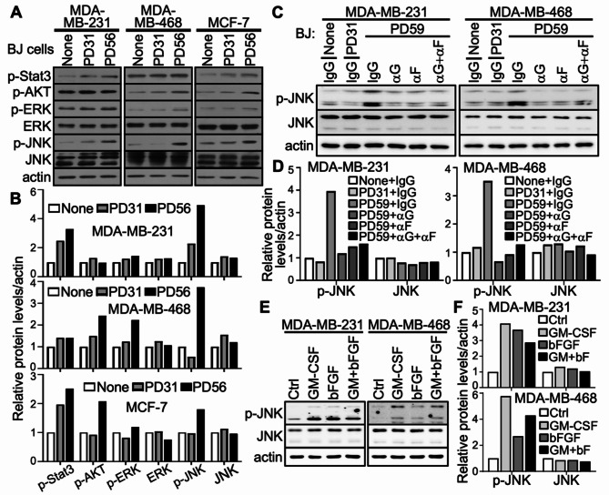
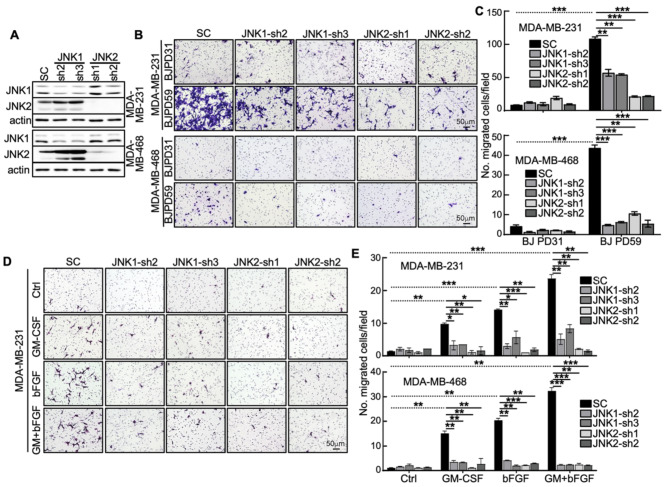
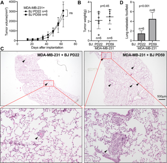
Results: Our results indicate that senescent cells stimulated the migration of breast cancer cells through secretion of GM-CSF and bFGF, which in turn induced activation of the JNK pathway in cancer cells. More importantly, senescent cells promoted breast cancer metastasis, with a minimum effect on the primary tumor growth, in orthotopic xenograft mouse models.

Conclusions: These results have revealed an additional mechanism by which senescent cells promote tumor cell metastasis and tumor progression, and will potentially lead to identification of novel targets for cancer therapies that suppress metastasis, the major cause of cancer mortality.

Keywords: Breast cancer; GM-CSF; JNK; Metastasis; Migration; Senescence; bFGF.


Conflict of interest statement

The authors declare no competing interests.

Figures

Fig. 1
Fig. 1
Senescent BJ cells promote the motility of breast cancer cells (A) Schematic diagram of the co-culture system in which young or senescent BJ cells and breast cancer cells were seeded in the lower and upper chambers, respectively, of the transwells. The 2 chambers were separated by a membrane with 8-micron pores. Migration of the breast cancer cells towards the lower chamber was measured. (B-C) Representative crystal violet-stained images of transwell migration of MDA-MB-231, MDA-MB-468 and MCF-7 cells co-cultured with medium (no cells or None) or young (PD34) or senescent (PD59) BJ cells for 12 h (B), and quantification of number of migrated cells per 20X field (mean ± SD, n = 3) (C). At least 5 randomly chosen 20X fields were counted for each of the triplicates. (D-E) Representative crystal violet-stained images of transwell migration of MDA-MB-231, MDA-MB-468 and MCF-7 cells co-cultured with medium only (no sup or None) or conditioned medium from young (PD32) or senescent (PD68) BJ cells for 16 h (D), and quantification of number of migrated cells per 20X field (mean ± SD, n = 3) (E). At least 5 randomly chosen 20X fields were counted for each of the triplicates. (F-G) Representative images of MDA-MB-231, MDA-MB-468 and MCF-7 cells immediately (0 h), 24 h (24 h, for MDA-MD-231 and MDA-MB-468) or 48 h (48 h, for MCF-7) after a scratch wound was made and co-cultured with medium only (no sup or None) or conditioned medium from young (PD26) or senescent (PD62) BJ cells (F), and quantification of the distance (µM) of the wound edges by ImageJ at 24 h (for MDA-MD-231 and MDA-MB-468) or 48 h (for MCF-7) (mean ± SD, n = 3) (G). (C, E, G) *** p < 0.001 between indicated groups in One-way ANOVA corrected for multiple comparisons using Tukey’s multiple comparison correction adjustment
Fig. 2
Fig. 2
Senescent cells secrete bFGF and GM-CSF that promote the motility of breast cancer cells (A) Relative mRNA levels (mean ± SD, n = 3) of GM-CSF, bFGF and CXCL1 in young (PD31) and senescent (PD62) BJ cells as determined by quantitative real time PCR analysis. (B) Relative protein levels (mean ± SD, n = 3) of GM-CSF, bFGF and CXCL1 in the conditioned medium from young (PD31) and senescent (PD62) BJ cells as determined by ELISA. (C-D) Representative crystal violet-stained images of transwell migration of MDA-MB-231, MDA-MB-468 and MCF-7 cells co-cultured with medium containing DMSO, 10 ng/ml of bFGF, 20 ng/ml of GM-CSF or both for 12 h (C), and quantification of number of migrated cells per field (mean ± SD, n = 3) (D). At least 5 randomly chosen 20X fields were counted for each of the triplicates. (E-F) Representative images of MDA-MB-231, MDA-MB-468 and MCF-7 cells immediately (0 h), 24 h (24 h, for MDA-MB-231 and MDA-MB-468) or 48 h (48 h, for MCF-7) after a scratch wound was made and co-cultured with medium containing DMSO, 10 ng/ml of bFGF, 20 ng/ml of GM-CSF or both (E), and quantification of the distance of the wound edges by ImageJ at 24 h (for MDA-MB-231 and MDA-MB-468) or 48 h (for MCF-7) (mean ± SD, n = 3) (F). (A-B, D, F) * p < 0.05, ** p < 0.01 and *** p < 0.001 vs. BJ-PD31 in unpaired, 2-sample t-tests (A-B) or between indicated groups in One-way ANOVA corrected for multiple comparisons using Tukey’s multiple comparison correction adjustment (D, F)
Fig. 3
Fig. 3
Senescent cells promote the migration of breast cancer cells via secreted bFGF and GM-CSF. (A-B) Representative crystal violet-stained images of transwell migration of MDA-MB-231, MDA-MB-468 and MCF-7 cells co-cultured with conditioned medium from young (PD29) or senescent (PD61) BJ cells, which were left untreated (None) or incubated with neutralizing antibodies against β-actin (1 ng/ml), bFGF (1 ng/ml), GM-CSF (5 ng/ml) or both bFGF and GM-CSF for 12 h (A), and quantification of number of migrated cells per field (mean ± SD, n = 3) (B). At least 5 randomly chosen 20X fields were counted for each of the triplicates (C-D) Representative crystal violet-stained images of transwell migration of MDA-MB-231, MDA-MB-468 and MCF-7 cells co-cultured with conditioned medium from young (PD29) or senescent (PD61) BJ cells transduced with shRNA control (SC) or shRNAs for bFGF or GM-CSF for 20 h (C), and quantification of number of migrated cells per field (mean ± SD, n = 3) (D). At least 5 randomly chosen 20X fields were counted for each of the triplicates. (B, D) ns, not significant; * p < 0.05; ** p < 0.01; and *** p < 0.001 between indicated groups in unpaired, 2-sample t tests (dotted lines) or One-way ANOVA corrected for multiple comparisons using Tukey’s multiple comparison correction adjustment (solid lines)
Fig. 4
Fig. 4
Senescent cells induce JNK activation via secreted GMCSF and bFGF (A-B) Western blotting analysis of the phosphorylation/activation status of Stat3, AKT, ERK and JNK in MDA-MB-231, MDA-MB-468 and MCF-7 breast cancer cell lines co-cultured with medium (None) or young (PD31) or senescent (PD56) BJ cells. (C-D) Western blotting analysis of phosphorylated/activated JNK in MDA-MB-231 and MDA-MB-468 breast cancer cell lines co-cultured with medium (None) or young (PD31) or senescent (PD59) BJ cells, which were incubated with neutralizing antibodies against IgG, GM-CSF (αG, 5 ng/ml), bFGF (αF, 1 ng/ml), or both bFGF and GM-CSF (αG +  αF). (E-F) Western blotting analysis of phosphorylated/activated JNK in MDA-MB-231 and MDA-MB-468 breast cancer cell lines treated with GM-CSF, bFGF or both. (B, D, F) Quantification of the Western blot results presented in A, C and E, respectively, showing expression levels of indicated proteins relative to None (B), None + IgG (D) or Ctrl (F). Densitometric quantification of bands in the Western blots was performed by ImageJ. The signals were normalized to that of actin
Fig. 5
Fig. 5
JNK mediates the stimulation of breast cancer migration by senescent cell-secreted GM-CSF and bFGF (A) Western blotting analysis showing knockdown specificity and efficiency of JNK1 and JNK2 shRNAs in MDA-MB-231 and MDA-MB-468 cells. (B-C) Representative crystal violet-stained images of transwell migration of MDA-MB-231 and MDA-MB-468 cells co-cultured with young (PD31) or senescent (PD59) BJ cells transduced with shRNA control (SC) or shRNAs for JNK1 or JNK2 for 12 h (C), and quantification of number of migrated cells per field (mean ± SD, n = 3) (D). At least 5 randomly chosen 20X fields were counted for each of the triplicates. (D-E) Representative crystal violet-stained images of transwell migration of MDA-MB-231 cells transduced with shRNAs for JNK1 or JNK2 or shRNA control (SC) and treated with vehicle control, 20 ng/ml of GM-CSF, 10 ng/ml of bFGF or both for 12 h (D), and quantification of number per field (mean ± SD, n = 3) of migrated MDA-MB-231 and MDA-MB-468 cells transduced with shRNAs for JNK1 or JNK2 or shRNA control (SC) and treated with vehicle control, 20 ng/ml of GM-CSF, 10 ng/ml of bFGF or both for 13 h (E). At least 5 randomly chosen 20X fields were counted for each of the triplicates. (C, E) **p < 0.01 and ***p < 0.001 between indicated groups in unpaired, 2-sample t tests (dotted lines) or One-way ANOVA corrected for multiple comparisons using Tukey’s multiple comparison correction adjustment (solid lines)
Fig. 6
Fig. 6
Senescent cells promote breast cancer metastasis in xenograft tumor models. 5 × 105 of MDA-MB-231 cells and 5 × 105 of young (PD22) or senescent (PD59) BJ cells were co-injected into the mammary fat pads of 6–8-week-old nude mice. Tumor sizes were measured weekly over 9 weeks. Tumor growth curves were plotted (A). Upon sacrifice, tumors were removed and weighted (B), and lung sections were stained by hematoxylin and eosin, photographed (C) and quantified for the number of metastatic foci (indicated by arrows) (D). In (C), images of the representative lung sections with metastatic foci (indicated by arrows) are shown in the top panels, and one metastatic focus (indicated by red boxes) from each section is magnified and shown in the bottom panels. In (D), for each mouse among the total of 6 in each group, the number of metastatic foci were counted in 5 randomly chosen 2X fields in a section containing all 5 lung lubes, and the number of metastatic foci per field was calculated. (A-B, D) Values are mean ± SD, n = 6 mice per group. p values are from unpaired, 2-sample t tests. ns, not significant

Similar articles

Cited by

  • The chemotherapy-induced senescence-associated secretome promotes cell detachment and metastatic dissemination through metabolic reprogramming.
    Cole AR, Buj R, Uboveja A, Levasseur E, Wang H, Kedziora KM, Chatoff A, Huacachino AA, Marcinkiewicz MM, Amalric A, Yang B, Tangudu NK, Danielson J, Elwah A, White S, Li D, Wallace CT, Lazure F, Elishaev E, Borho L, Jazwinska DE, Laird MS, Atiya H, Bitler BG, Dangaj D, Coffman LG, Tseng G, Oesterreich S, Gomes AP, Gurkar AU, Schopfer FJ, Modugno F, Watkins SC, Zervantonakis IK, Stallaert W, Hempel N, Snyder NW, Aird KM. Cole AR, et al. bioRxiv [Preprint]. 2025 Aug 12:2023.12.02.569652. doi: 10.1101/2023.12.02.569652. bioRxiv. 2025. PMID: 40832218 Free PMC article. Preprint.

References

    1. Parkin DM. Global cancer statistics in the year 2000. Lancet Oncol. 2001;2(9):533–43. - PubMed
    1. Coppe JP, Kauser K, Campisi J, Beausejour CM. Secretion of vascular endothelial growth factor by primary human fibroblasts at senescence. J Biol Chem. 2006;281(40):29568–74. - PubMed
    1. Laberge RM, Awad P, Campisi J, Desprez PY. Epithelial-mesenchymal transition induced by senescent fibroblasts. Cancer Microenvironment: Official J Int Cancer Microenvironment Soc. 2012;5(1):39–44. - PMC - PubMed
    1. Campisi J. Cancer, aging and cellular senescence. vivo. 2000;14(1):183–8. - PubMed
    1. Krtolica A, Parrinello S, Lockett S, Desprez PY, Campisi J. Senescent fibroblasts promote epithelial cell growth and tumorigenesis: a link between cancer and aging. Proc Natl Acad Sci USA. 2001;98(21):12072–7. - PMC - PubMed

Substances