

Outstanding increase in tumor-to-background ratio over time allows tumor localization by [89Zr]Zr-PSMA-617 PET/CT in early biochemical recurrence of prostate cancer
- PMID: 39375762
- PMCID: PMC11457487
- DOI: 10.1186/s40644-024-00778-5
Outstanding increase in tumor-to-background ratio over time allows tumor localization by [89Zr]Zr-PSMA-617 PET/CT in early biochemical recurrence of prostate cancer
Abstract
Background: Positron emission tomography/computed tomography (PET/CT) using prostate-specific membrane antigen (PSMA)-targeted radiotracers labeled with zirconium-89 (89Zr; half-life ~ 78.41 h) showed promise in localizing biochemical recurrence of prostate cancer (BCR) in pilot studies.
Methods: Retrospective analysis of 38 consecutive men with BCR (median [minimum-maximum] prostate-specific antigen 0.52 (0.12-2.50 ng/mL) undergoing [89Zr]Zr-PSMA-617 PET/CT post-negative [68Ga]Ga-PSMA-11 PET/CT. PET/CT acquisition 1-h, 24-h, and 48-h post-injection of a median (minimum-maximum) [89Zr]Zr-PSMA-617 tracer activity of 123 (84-166) MBq.
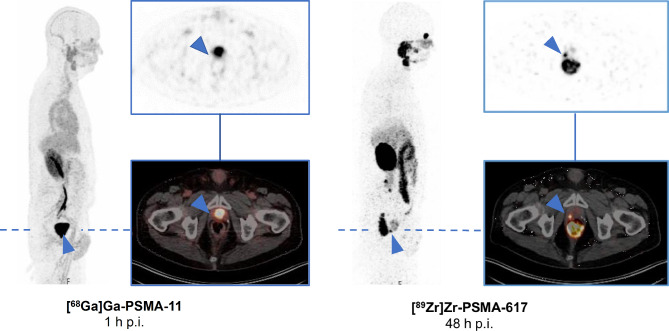
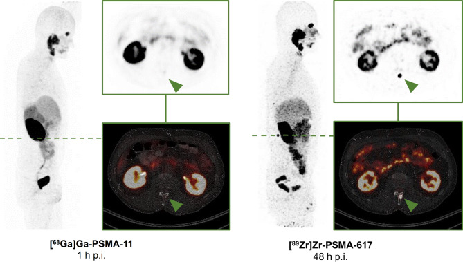
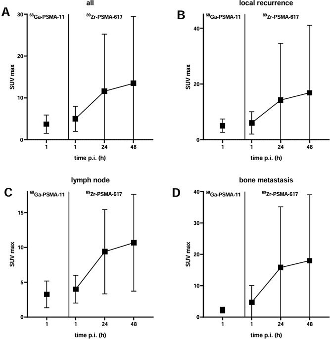
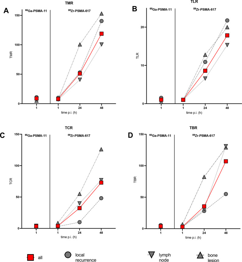
Results: [89Zr]Zr-PSMA-617 PET/CT detected altogether 57 lesions: 18 local recurrences, 33 lymph node metastases, 6 bone metastases in 30/38 men with BCR (78%) and prior negative conventional PSMA PET/CT. Lesion uptake significantly increased from 1-h to 24-h and, in a majority of cases, from 24-h to 48-h. Tumor-to-background ratios significantly increased over time, with absolute increases of 100 or more. No side effects were noted. After [89Zr]Zr-PSMA-617 PET/CT-based treatment, prostate-specific antigen concentration decreased in all patients, becoming undetectable in a third of patients.
Limitations: retrospective, single center design; infrequent histopathological and imaging verification.
Conclusion: This large series provides further evidence that [89Zr]Zr-PSMA-617 PET/CT is a beneficial imaging modality to localize early BCR. A remarkable increase in tumor-to-background ratio over time allows localization of tumor unidentified on conventional PSMA PET/CT.
Keywords: Biochemical recurrence; Localization; Positron emission tomography/Computed tomography (PET/CT); Prostate cancer; Prostate-specific membrane antigen (PSMA); Zirconium-89 (89Zr).
© 2024. The Author(s).
Conflict of interest statement
The authors declare no competing interests.
Figures


References
-
- Grunig H, Maurer A, Thali Y, Kovacs Z, Strobel K, Burger IA, et al. Focal unspecific bone uptake on [18F]-PSMA-1007 PET: a multicenter retrospective evaluation of the distribution, frequency, and quantitative parameters of a potential pitfall in prostate cancer imaging. Eur J Nucl Med Mol Imaging. 2021;48:4483–94. - PMC - PubMed
-
- Bagguley D, Ong S, Buteau JP, Koschel S, Dhiantravan N, Hofman MS, et al. Role of PSMA PET/CT imaging in the diagnosis, staging and restaging of prostate cancer. Future Oncol. 2021;17:2225–41. - PubMed
-
- Perera M, Papa N, Roberts M, Williams M, Udovicich C, Vela I, et al. Gallium-68 prostate-specific membrane Antigen Positron Emission Tomography in Advanced prostate Cancer-updated diagnostic utility, sensitivity, specificity, and distribution of prostate-specific membrane Antigen-avid lesions: a systematic review and Meta-analysis. Eur Urol. 2020;77:403–17. - PubMed
MeSH terms
Substances
LinkOut - more resources
Full Text Sources
Medical
Miscellaneous
