Review
. 2024 Oct 7;28(1):328.
doi: 10.1186/s13054-024-05102-y.

Nuts and bolts of lung ultrasound: utility, scanning techniques, protocols, and findings in common pathologies

Affiliations
Review

Nuts and bolts of lung ultrasound: utility, scanning techniques, protocols, and findings in common pathologies

Michael Beshara et al. Crit Care. .

Abstract

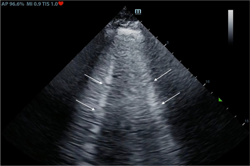
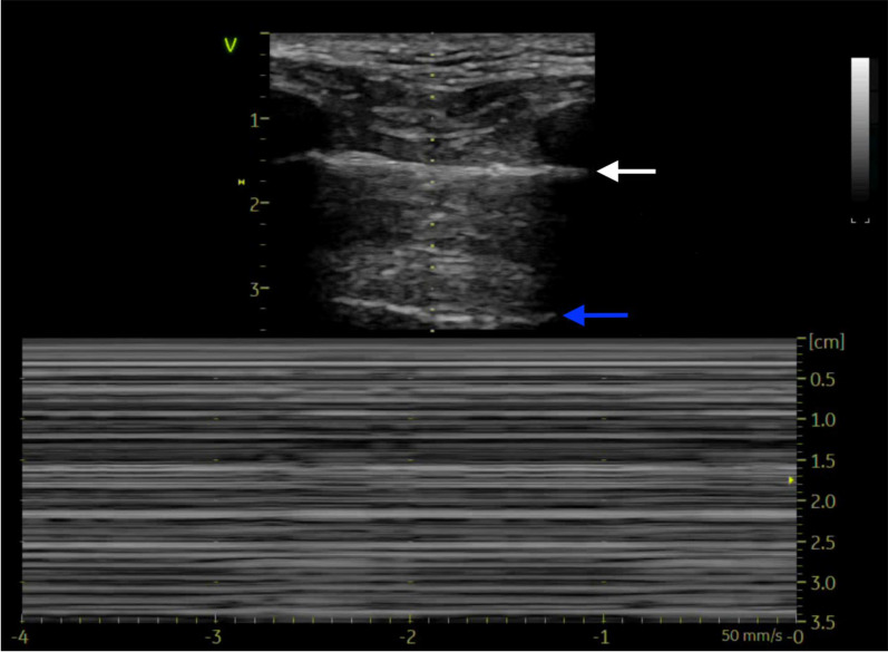
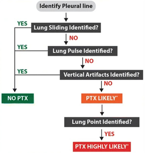
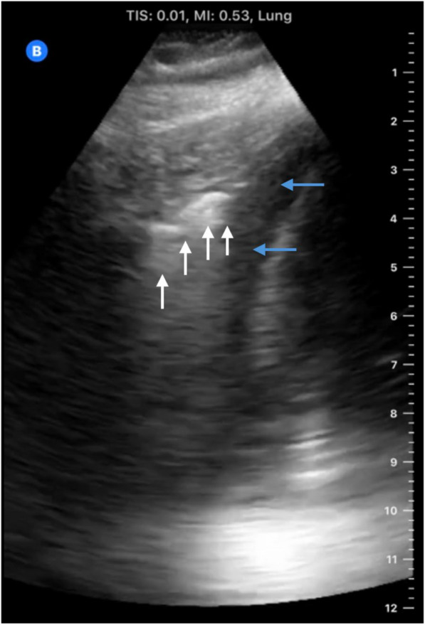
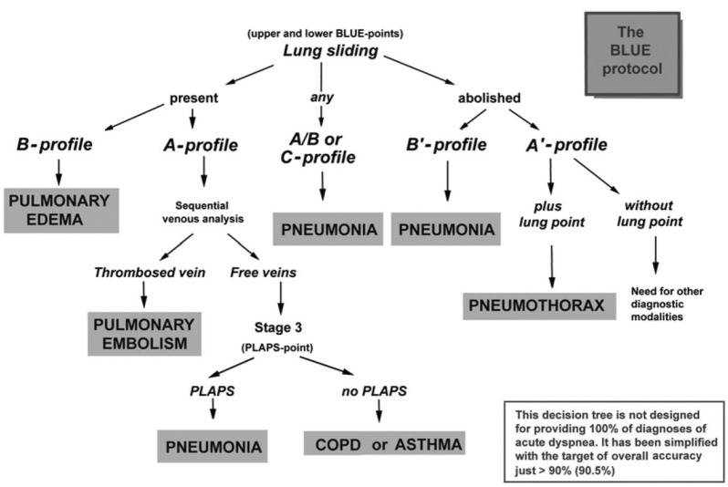
Point of Care ultrasound (POCUS) of the lungs, also known as lung ultrasound (LUS), has emerged as a technique that allows for the diagnosis of many respiratory pathologies with greater accuracy and speed compared to conventional techniques such as chest x-ray and auscultation. The goal of this narrative review is to provide a simple and practical approach to LUS for critical care, pulmonary, and anesthesia providers, as well as respiratory therapists and other health care providers to be able to implement this technique into their clinical practice. In this review, we will discuss the basic physics of LUS, provide a hands-on scanning technique, describe LUS findings seen in normal and pathological conditions (such as mainstem intubation, pneumothorax, atelectasis, pneumonia, aspiration, COPD exacerbation, cardiogenic pulmonary edema, ARDS, and pleural effusion) and also review the training necessary to achieve competence in LUS.

Keywords: ARDS; Anesthesia; Aspiration; Atelectasis; COPD exacerbation; Cardiogenic pulmonary edema; Critical care; Critical care ultrasound; LUS; Lung ultrasound; Lung ultrasound training; Mainstem intubation; POCUS; POCUS competency; POCUS training; Pleural effusion; Pneumonia; Pneumothorax; Point of care ultrasound; Pulmonary; Ultrasound; Ultrasound competency; Ultrasound training.


Conflict of interest statement

None.

Figures

Fig. 1
Fig. 1
B-lines (white arrows) which are vertical hyperechoic lines that begin at the pleural line and extend all the way down to the bottom of the ultrasound screen
Fig. 2
Fig. 2
Top panel is a 2D image of pneumothorax depicting the pleural line (white arrow) and A-line (blue arrow) with no B-lines. Bottom panel is an M-mode image in pneumothorax showing stratosphere or Barcode sign
Fig. 3
Fig. 3
Simple Algorithm for diagnosis of Pneumothorax. Reproduced with permission from Dr. Goffi [62]
Fig. 4
Fig. 4
Consolidated lung tissue. The irregular border “Shred sign” between the consolidated and normal lung tissue (white arrows). The lung here is surrounded by an pleural effusion (blue lines)
Fig. 5
Fig. 5
The Bedside Lung Ultrasound in Emergency (BLUE) Protocol for patients with acute respiratory failure. A-profile: Lung sliding + A-lines, B-profile: Lung sliding + B-lines, A/B-profile: A-profile in one lung and B-profile in the other, C-profile: Anterior lung consolidation, A’-profile: A profile + negative lung sliding, B’-profile: B profile + negative lung sliding. Reproduced with permission from Dr. Lichtenstein [7, 25]

References

    1. Kendall JL, Hoffenberg SR, Smith RS. History of emergency and critical care ultrasound: the evolution of a new imaging paradigm. Crit Care Med. 2007. 10.1097/01.CCM.0000260623.38982.83. - PubMed
    1. Rice JA, Brewer J, Speaks T, Choi C, Lahsaei P, Romito BT. The POCUS consult: how point of care ultrasound helps guide medical decision making. Int J Gen Med. 2021;14:9789–806. 10.2147/IJGM.S339476. - PMC - PubMed
    1. Arnold MJ, Jonas CE, Carter RE. Point-of-care ultrasonography. Am Fam Physician. 2020;101(5):275–85. - PubMed
    1. Soldati G, Demi M, Smargiassi A, Inchingolo R, Demi L. The role of ultrasound lung artifacts in the diagnosis of respiratory diseases. Expert Rev Respir Med. 2019;13(2):163–72. 10.1080/17476348.2019.1565997. - PubMed
    1. Convissar DL, Gibson LE, Berra L, Bittner EA, Chang MG. Application of lung ultrasound during the COVID-19 pandemic: a narrative review. Anesth Analg. 2020;131(2):345–50. 10.1213/ANE.0000000000004929. - PMC - PubMed

MeSH terms

LinkOut - more resources