. 2024 Dec;20(51):e2405927.
doi: 10.1002/smll.202405927. Epub 2024 Oct 7.

Microneedles with Implantable Tip-Accumulated Therapeutics for the Long-Term Management of Psoriasis

Affiliations

Microneedles with Implantable Tip-Accumulated Therapeutics for the Long-Term Management of Psoriasis

Fatma Moawad et al. Small. 2024 Dec.

Abstract

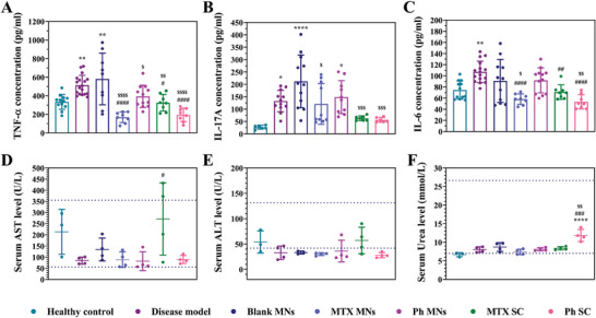
Methotrexate is successfully used as the gold standard for managing moderate-to-severe psoriasis. However, the low bioavailability and short half-life of the oral pills and the invasiveness of the parenteral injections make these suboptimal therapeutic options. Microneedles, bridging the advantages of the former forms, are successfully used to deliver methotrexate for different therapeutic purposes. However, the utilized dissolving microneedles demand frequent administration, potentially compromising patients' compliance. Additionally, the high toxicity of methotrexate prompts a quest for safer alternatives. Phloretin, a natural compound with confirmed antipsoriatic potential, emerges as a promising candidate. Herein, microneedle patches with separable, slow-degrading tips are developed for the sustained delivery of methotrexate and phloretin, as a comprehensive solution for long-term psoriasis management. Both compounds are individually loaded at varying doses and display sustained-release profiles. The developed microneedle patches demonstrate high mechanical strength, favorable drug delivery efficiency, and remarkable antipsoriatic potential both in vitro in keratinocytes and in vivo in a psoriasis mouse model. Comparative analysis with two subcutaneous injections reveals a similar antipsoriatic efficacy with a single patch of either compound, with prominent phloretin safety. Therefore, the developed patches present a superior alternative to methotrexate's current marketed forms and provide a viable alternative (phloretin) with comparable antipsoriatic efficacy and higher safety.

Keywords: PLGA; methotrexate; microneedles; phloretin; psoriasis; sustained release.


Conflict of interest statement

The authors declare no conflict of interest.

Figures

Figure 1
Figure 1
General characterization of the PLGA‐tipped MN patches. A) Top‐view fluorescence (i) side‐view fluorescence (iii), and side‐view bright‐field (iv) images of an MN patch with Cy5‐loaded PLGA tips, and bright‐field image (ii) of the PLGA tips after the dissolution of one patch in water (scale bar = 1 mm for (i), 100 µm for (ii), and 500 µm for (iii, iv)). B) Top‐view bright‐field image and C) SEM micrograph of a blank PLGA‐tipped MN patch (scale bar = 1 mm and 300 µm, respectively). D) Force‐displacement curve of the blank PLGA‐tipped MNs. E) Side‐view bright‐field images of the blank PLGA‐tipped MNs before (i) and after (ii) mechanical testing (scale bar = 200 µm and is applied to the other image in the same figure). F) SEM micrograph of the blank PLGA‐tipped MNs after mechanical testing (scale bar = 300 µm). G) Penetration, implantation, and drug delivery profile of Cy5‐loaded PLGA‐tipped MNs. (H) Bright‐field (i) and fluorescence (ii) images of excised porcine skin following the application of an MN patch and bright‐field images of an MN patch before (iii) and after (iv) application into the skin for 5 min (scale bars = 1 mm and is applied to the other image in the same raw). I) H&E‐stained histology section of the skin showing a penetration site of MN (scale bar = 100 µm). (Mean ± SD; n = 4–6).
Figure 2
Figure 2
General characterization of MTX and Ph‐loaded PLGA‐tipped MNs, as well as their individual components. A) bright‐field images (i, ii) and SEM micrographs (v, vi) of MTX (i, v) and Ph‐ (ii, vi) loaded MNs (scale bar = 1 mm for (i, ii) and scale bar = 300 µm for (v, vi)), and SEM micrographs of raw MTX (iii) and Ph (iv) (scale bar = 5 and 50 µm, respectively). B) Force‐displacement curves of blank, MTX‐, and Ph‐loaded MNs. C) Bright‐field images of MTX (i) and Ph‐ (ii) loaded MNs following mechanical testing (scale bar = 200 µm). D) XRD spectra of MTX and Ph‐loaded MNs and their individual raw components. E) DSC thermograms of MTX and Ph‐loaded MNs and their individual raw components. (Mean ± SD; n = 4–6).
Figure 3
Figure 3
Loading, release profiles, and in vitro antiproliferative activity of MTX and Ph‐loaded PLGA‐tipped MNs. A) MN content of MTX and Ph at different initial concentrations of each compound (n = 4). B) In vitro release profile of MTX and Ph from the PLGA‐tipped MNs (n = 5). (C,D) Viability percentages of HaCat cells treated with different concentrations of MTX and Ph, respectively, extracted from fresh and stored MNs, and compared to the standard solution (SS) of each compound, blank MNs, and negative controls (n = 5 wells per each condition). Mean ± SD.
Figure 4
Figure 4
In vivo efficacy and safety of MTX and Ph‐loaded PLGA‐tipped MNs: impact of the therapy on the phenotypic manifestations of psoriasis. A) Schematic diagram of the experimental protocol of the in vivo efficacy study. B) Representative macroscopic images and C) total PASI score of mice dorsal lesions from the different in vivo groups throughout the experimental period. D) Change in mice body weight (%) throughout the whole experimental period. E) Spleen mass index (%) of the different in vivo groups. Asterisks (*) mark the statistical difference between the different groups and the healthy control, and hashes (#) mark the statistical difference between the different groups and the disease model. (Mean ± SD; n = 3–4).
Figure 5
Figure 5
In vivo efficacy of MTX and Ph‐loaded PLGA‐tipped MNs: impact of the therapy on the expression level of key markers of psoriasis. A) Representative images of H&E‐stained sections of mice dorsal skin and E) the corresponding calculated epidermal thicknesses using the Zen 3.1 software (15–20 measurements were recorded from the skin of each mouse and the averages were calculated, with the epidermal protrusions taken into consideration during measurement). (B,C) Representative images of immunohistochemically stained skin sections for Ki67+ and CD3+ cells, respectively, and the corresponding quantitative analysis (F), G) calculated using the ImageJ software (three images were analyzed from the skin of each mouse). D) Representative images of H&E‐stained sections of the spleens from the different in vivo groups. Asterisks (*) mark the statistical difference between the different groups and the healthy control, hashes (#) mark the statistical difference between the different groups and the disease model, and dollar signs ($) mark the statistical difference between the different groups and the blank MN group. The scale bar in each image is applied to other images in the same row. (Mean ± SD; n = 3–4).
Figure 6
Figure 6
In vivo efficacy and safety of MTX and Ph‐loaded PLGA‐tipped MNs: impact of the therapy on the expression level of primary cytokine targets of psoriasis and blood biochemical analysis. The concentration of A) TNF‐α, B) IL‐17A, and C) IL‐6 in the skin homogenate of the different in vivo groups, detected via ELISA. Serum levels of D) AST, E) ALT, and F) urea, the dotted lines represent the reference ranges for the Balb/C mice. Asterisks (*) mark the statistical difference between the different groups and the healthy control, and hashes (#) mark the statistical difference between the different groups and the disease model. (Mean ± SD; n = 3–4).

Similar articles

Cited by

References

    1. Griffiths C. E., Barker J. N., Lancet 2007, 370, 263. - PubMed
    1. a) Greb J. E., Goldminz A. M., Elder J. T., Lebwohl M. G., Gladman D. D., Wu J. J., Mehta N. N., Finlay A. Y., Gottlieb A. B., Nat. Rev. Dis. Primers 2016, 2, 16082; - PubMed
    2. b) Menter A., Griffiths C. E., Lancet 2007, 370, 272. - PubMed
    1. a) Chen Z., Br. J. Dermatol. 2018, 179, 16908;
    2. b) Law J. H., Koo B., Koo J. Y., Psoriasis Forum 2008, 14, 17.
    1. Cronstein B. N., Aune T. M., Nat. Rev. Rheumatol. 2020, 16, 145. - PubMed
    1. Negrei C., Boda D., An Interdisciplinary Approach to Psoriasis, 2017, IntechOpen, London, p. 211.

Publication types

LinkOut - more resources