. 2024 Oct 8:15910199241272596.
doi: 10.1177/15910199241272596. Online ahead of print.

Recognition of dural to pial supply in high-grade dural arteriovenous fistula: A technical note

Affiliations

Recognition of dural to pial supply in high-grade dural arteriovenous fistula: A technical note

Shigeta Miyake et al. Interv Neuroradiol. .

Abstract

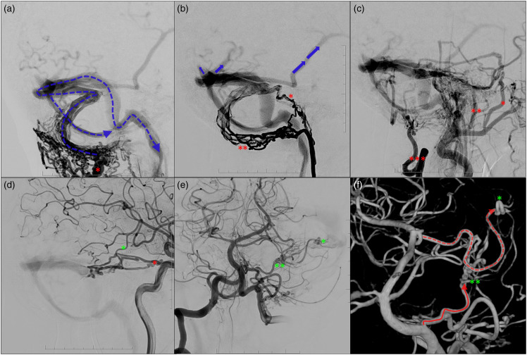
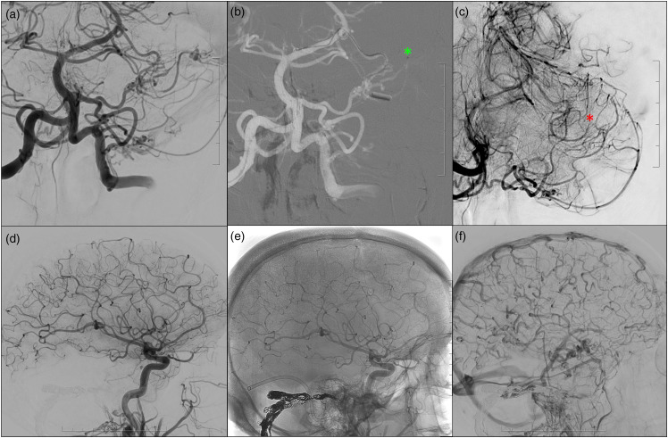
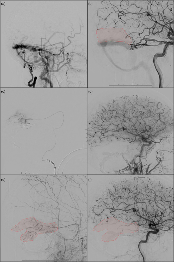
High-grade dural arteriovenous fistulas (DAVFs) are known to demonstrate classical dural supply and can demonstrate pre-existing dural supply and 'pure' arterial supply from pial branches. The latter two are examples of congenital versus acquired pial to dural shunting, respectively. We describe the recognition of dural to pial supply during combined transarterial and transvenous embolization of a high-grade DAVF with holocephalic venous reflux, stressing the importance of careful assessment of this condition with micro catheter injections.

Keywords: Dural arteriovenous fistula; VEGF; dural supply; endovascular treatment; pial supply.


Conflict of interest statement

Declaration of conflicting interestsThe author(s) declared the following potential conflicts of interest with respect to the research, authorship, and/or publication of this article: EH is consultant for Medtronic.

Figures

Figure 1.
Figure 1.
DSA shows a multi-hole high-grade DAVF located at the left distal transverse sinus, continuing along an adjacent parasinus in the posterior fossa. There is extensive transosseous supply from the left deep cervical artery (a; red asterisk), originating from the left subclavian artery, classical dural supply from left external carotid artery branches including the neuromeningeal trunk of the left ascending pharyngeal artery (b; single and double red asterisks), the left occipital artery (c; triple red asterisks) and left superficial temporal artery, left posterior auricular artery, left deep temporal artery and left distal internal maxillary artery branches (c). There was supply from the left internal carotid artery lateral and medial tentorial branches (d; red asterisk). There was supply from pre-existing dural to pial branches: the posterior meningeal arteries from the right and left postero-inferior cerebrellar artery (PICA; e) and left antero-inferior cerebellar artery (or subarcuate artery; e; double green asterisks). There was ‘pure’ arterial supply from the left P2 posterior cerebral artery (e; single green asterisk) and smaller more distal right MCA branches (d; single green asterisk). There was retrograde drainage into the ipsilateral dural venous system (a), with dural venous reflux into the distal superior sagittal sinus and straight sinus and cortical venous reflux to the left cerebral and cerebellar hemisphere, including the contralateral vein of Labbe (b).
Figure 2.
Figure 2.
Prior to transvenous coil embolization via a contralateral transjugular approach, the ‘pure’ arterial supply of the left distal PCA branch was obliterated via superselective embolization with 50% Gluebran: 50% Lipiodol through a 1.2 Magic flow-directed microcatheter (BALT neurovascular), pre embolization (b; green asterisk) and post embolization (c; red asterisk). The pre-existing dural supply from the left subarcuate artery was left untouched. Subsequently, the affected parasinus in the posterior fossa and right distal transverse sinus were occluded with a dual-microcatheter technique with coils, with complete occlusion of the DAVF after embolization (d, subtracted image; e, unsubtracted images demonstrating the coil skeleton). There is global slow flow in cortical veins with normalized antegrade flow in the contralateral vein of Labbe on post embolization angiogram (f).
Figure 3.
Figure 3.
The pre-treatment external carotid injection shows numerous classical feeders, but no cortical supply of the MMA (a). On the other hand, the internal carotid artery (ICA) injection showed an avascular area distal to the pial supply of the MCA branch (b, red territory). A superselective injection of the petrosquamosal branch of the right MMA was performed to confirm closure of the DAVF during the procedure. Microinjection demonstrated dural to pial supply to a large area of the right posterior middle temporal gyrus (c) and possibly a smaller area of the left superior cerebellar artery territory. ICA injection performed after closure of the DAVF showed slow flow of the distal MCA where the pial supply was located, and the distal parenchyma was replaced by blood flow from the MMA. The dural to pial supply was confirmed on three-month follow-up cerebral angiography, with established supply from the MMA to the right temporal lobe (e and f).

Similar articles

Cited by

References

    1. van Dijk JMC, terBrugge KG, Willinsky RA, et al. Clinical course of cranial dural arteriovenous fistulas with long-term persistent cortical venous reflux. Stroke 2002; 33: 1233–1236. - PubMed
    1. Osada T, Krings T. Intracranial dural arteriovenous fistulas with pial arterial supply. Neurosurgery 2019; 84: 104–115. - PubMed
    1. Brinjikji W, Cloft HJ, Lanzino G. Clinical, angiographic, and treatment characteristics of cranial dural arteriovenous fistulas with pial arterial supply. J Neurointerv Surg 2021; 13: 331–335. 20200629. - PubMed
    1. Wu Q, Zhang X-S, Wang H-D, et al. Onyx embolization for tentorial dural arteriovenous fistula with pial arterial supply: case series and analysis of complications. World Neurosurg 2016; 92: 58–64. 20160420. - PubMed
    1. Sato K, Matsumoto Y, Endo H, et al. A hemorrhagic complication after onyx embolization of a tentorial dural arteriovenous fistula: a caution about subdural extension with pial arterial supply. Interv Neuroradiol 2017; 23: 307–312. 20170101. - PMC - PubMed

LinkOut - more resources