T2 MRI visible perivascular spaces in Parkinson's disease: clinical significance and association with polysomnography measured sleep
- PMID: 39377177
- PMCID: PMC11725513
- DOI: 10.1093/sleep/zsae233
T2 MRI visible perivascular spaces in Parkinson's disease: clinical significance and association with polysomnography measured sleep
Abstract
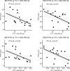
Poor sleep quality might contribute to the risk and progression of neurodegenerative disorders via deficient cerebral waste clearance functions during sleep. In this retrospective cross-sectional study, we explore the link between enlarged perivascular spaces (PVS), a putative marker of sleep-dependent glymphatic clearance, with sleep quality and motor symptoms in patients with Parkinson's disease (PD). T2-weighted magnetic resonance imaging (MRI) images of 20 patients and 17 healthy control participants were estimated visually for PVS in the basal ganglia (BG) and centrum semiovale (CSO). The patient group additionally underwent a single-night polysomnography. Readouts included polysomnographic sleep features and slow-wave activity (SWA), a quantitative EEG marker of sleep depth. Associations between PVS counts, PD symptoms (MDS-UPDRS scores), and sleep parameters were evaluated using correlation and regression analyses. Intra- and inter-rater reproducibility was assessed with weighted Cohen`s kappa coefficient. BG and CSO PVS counts in both patients and controls did not differ significantly between groups. In patients, PVS in both brain regions was negatively associated with SWA (1-2 Hz; BG: r(15) = -.58, padj = .015 and CSO: r(15) = -.6, padj = .015). Basal ganglia PVS counts were positively associated with motor symptoms of daily living (IRR = 1.05, CI [1.01, 1.09], p = .007, padj = .026) and antidepressant use (IRR = 1.37, CI [1.05, 1.80], p = .021, padj = .043) after controlling for age. Centrum Semiovale PVS counts in patients were positively associated with a diagnosis of REM sleep behavior disorder (IRR = 1.39, CI [1.06, 1.84], p = .018, padj = .11). These results add to evidence that sleep deterioration may play a role in impairing glymphatic clearance via altered perivascular function, potentially contributing to disease severity in PD patients.
Keywords: Parkinson; glymphatic; magnetic resonance imaging; neurodegeneration; perivascular spaces; sleep; slow-wave sleep; waste clearance.
© The Author(s) 2024. Published by Oxford University Press on behalf of Sleep Research Society.
Figures


Comment in
-
Glymphatic system, sleep, and Parkinson's disease: interconnections, research opportunities, and potential for disease modification.Sleep. 2025 Jan 13;48(1):zsae251. doi: 10.1093/sleep/zsae251. Sleep. 2025. PMID: 39450429 Free PMC article. No abstract available.
References
-
- Owens T, Bechmann I, Engelhardt B.. Perivascular spaces and the two steps to neuroinflammation. J Neuropathol Exp Neurol. 2008;67(12):1113–1121. doi: https://doi.org/10.1097/NEN.0b013e31818f9ca8 - DOI - PubMed
-
- Iliff JJ, Wang M, Liao Y, et al.A Paravascular Pathway facilitates CSF flow through the brain parenchyma and the clearance of interstitial solutes, including Amyloid β. Sci Transl Med. 2012;4(147):147ra111. doi: https://doi.org/10.1126/scitranslmed.3003748 - DOI - PMC - PubMed
-
- Xie L, Kang H, Xu Q, et al.Sleep drives metabolite clearance from the adult brain. Science. 2013;342(6156):373–377. doi: https://doi.org/10.1126/science.1241224 - DOI - PMC - PubMed
-
- Wardlaw JM, Benveniste H, Nedergaard M, et al.; colleagues from the Fondation Leducq Transatlantic Network of Excellence on the Role of the Perivascular Space in Cerebral Small Vessel Disease. Perivascular spaces in the brain: anatomy, physiology and pathology. Nat Rev Neurol. 2020;16(3):137–153. doi: https://doi.org/10.1038/s41582-020-0312-z - DOI - PubMed
-
- Ramirez J, Berezuk C, McNeely AA, Gao F, McLaurin J, Black SE.. Imaging the perivascular space as a potential biomarker of neurovascular and neurodegenerative diseases. Cell Mol Neurobiol. 2016;36(2):289–299. doi: https://doi.org/10.1007/s10571-016-0343-6 - DOI - PMC - PubMed
MeSH terms
Grants and funding
LinkOut - more resources
Full Text Sources
Medical
Research Materials
Miscellaneous

