. 2024 Oct 15;13(20):e034690.
doi: 10.1161/JAHA.124.034690. Epub 2024 Oct 8.

Defining Cardiomyocyte Repolarization Response to Pharmacotherapy in Long-QT Syndrome Type 3

Affiliations

Defining Cardiomyocyte Repolarization Response to Pharmacotherapy in Long-QT Syndrome Type 3

Ning Ge et al. J Am Heart Assoc. .

Abstract

Background: Long-QT syndrome is a primary cardiac ion channelopathy predisposing a patient to ventricular arrhythmia through delayed repolarization on the resting ECG. We aimed to establish a patient-specific, human induced pluripotent stem cell (hiPSC)-derived cardiomyocytes model of long-QT syndrome type 3 (LQT3) using clustered regularly interspaced palindromic repeats (CRISPR/Cas9), for disease modeling and drug challenge.

Methods and results: HiPSCs were generated from a patient with LQT3 harboring an SCN5A pathogenic variant (c.1231G>A; p.Val411Met), and an unrelated healthy control. The same SCN5A pathogenic variant was engineered into the background healthy control hiPSCs via CRISPR/Cas9 gene editing to generate a second disease model of LQT3 for comparison with an isogenic control. All 3 hiPSC lines were differentiated into cardiomyocytes. Both the patient-derived LQT3 (SCN5A+/-) and genetically engineered LQT3 (SCN5A+/-) hiPSC-derived cardiomyocytes showed significantly prolonged cardiomyocyte repolarization compared with the healthy control. Mexiletine, a cardiac voltage-gated sodium channel (NaV1.5) blocker, shortened repolarization in both patient-derived LQT3 and genetically engineered LQT3 hiPSC-derived cardiomyocytes, but had no effect in the control. Notably, calcium channel blockers nifedipine and verapamil showed a dose-dependent shortening of repolarization, rescuing the phenotype. Additionally, therapeutic drugs known to prolong the corrected QT in humans (ondansetron, clarithromycin, and sotalol) demonstrated this effect in vitro, but the LQT3 clones were not more disproportionately affected compared with the control.

Conclusions: We demonstrated that patient-derived and genetically engineered LQT3 hiPSC-derived cardiomyocytes faithfully recapitulate pathologic characteristics of LQT3. The clinical significance of such an in vitro model is in the exploration of novel therapeutic strategies, stratifying drug adverse reaction risk and potentially facilitating a more targeted, patient-specific approach in high-risk patients with LQT3.

Keywords: CRISPR/Cas9; SCN5A; human induced pluripotent stem cells; long‐QT syndrome; multielectrode array.


Figures

Figure 1
Figure 1. ECG and genotyping of a patient with LQT3 and healthy control.
A, ECGs from an unrelated healthy control and a patient with LQT3. Horizontal color bars (blue) represent QT intervals on control and patient ECGs. The patient with LQT3 (458 ms) displayed prolonged corrected QT interval compared with healthy control (362 ms). B, Sanger sequencing chromatograms of healthy control and patient with LQT3. C, Illustration showing the position of the Val411Met pathogenic variant in human cardiac voltage‐gated sodium channel protein (NaV1.5). See also Figure S1. hiPSCs indicates human induced pluripotent stem cells; LQT3, long‐QT syndrome type 3; LQTS, long‐QT syndrome; and QTc, corrected QT.
Figure 2
Figure 2. Generation and characterization of hiPSCs and hiPSC‐CMs.
A, Characterization of a patient with LQT3 hiPSC cl1. Representative images of hiPSC morphology and alkaline phosphatase staining on left‐sided panels; immunofluorescence staining of hiPSC clones showing positive expression of pluripotent stem cell markers OCT4 (POU class 5 homeobox 1), SOX2 (SRY‐box transcription factor 2), NANOG (Nanog homeobox), TRA‐1‐60 (Tumor‐related antigen 1‐60), TRA‐1‐81 (Tumor‐related antigen 1‐81), and SSEA4 (stage‐specific embryonic antigen 4) (Hoechst staining of nuclear DNA and merged images). Scale bar: 100 μm. All of the cell line data are in Figure S3A. B, Workflow of cardiac differentiation from hiPSCs to differentiated cardiomyocytes. C, Representative flow cytometry plots for expression of troponin T in control cl1, patient with LQT3 cl1, and genetically engineered LQT3 cl1 hiPSC‐CMs on day 14 of differentiation. All of the cell line data are in Figure S6B. D, Bar graph of the relative messenger RNA expression level of cardiomyocyte genes TNNT2 and sarcomere genes ACTN2 in comparison with the housekeeping gene GAPDH and normalized to the control cl1 on day 28 of differentiation (n=3). E, Representative images of beating cardiomyocytes for a patient with LQT3 cl1 hiPSC‐CMs on day 14 of differentiation, and immunocytochemical staining of ventricular cardiomyocyte marker MYL2 (MLC‐2v), cardiac marker troponin T, and sarcomere protein α‐actinin for a patient with LQT3 cl1 hiPSC‐CMs on day 28 of differentiation. Scale bar: 50 μm. Error bars represent SEM. See also Figures S3 and S6. (B) was created with Biorender.com. hiPSC‐CMs indicates human induced pluripotent stem cell‐derived cardiomyocytes; hiPSCs indicates human induced pluripotent stem cells; and LQT3, long‐QT syndrome type 3.
Figure 3
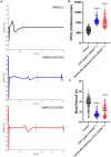
Figure 3. Electrophysiological properties of control, patient with LQT3, and genetically engineered LQT3 hiPSC‐CMs.
A, Representative field potential wave of healthy control (top, black), patient with LQT3 (middle, blue), and genetically engineered (induced) LQT3 (bottom, red) hiPSC‐CMs on day 28 of differentiation. B and C, FPDc and beat period from the averaged values of all 3 clones of cardiomyocytes derived from the healthy control, patient with LQT3, and genetically engineered LQT3 cell lines. N=240 per group. Statistical significance ****P<0.0001 patient with LQT3 and genetically engineered LQT3 vs healthy control group. Error bars represent SEM. See also Figure S7. FPDc indicates corrected field potential duration; hiPSC‐CMs, human induced pluripotent stem cell‐derived cardiomyocytes; and LQT3, long‐QT syndrome type 3.
Figure 4
Figure 4. hiPSC‐CMs in response to drug treatment.
FPDc values measured from representative hiPSC‐CMs clones of healthy control, patient with LQT3, and genetically engineered LQT3 cell lines following administration of increasing concentrations of mexiletine (A), E‐4031 (B), nifedipine (C), verapamil (D), ondansetron (E), clarithromycin (F), and sotalol (G). Normalized FPDc values to baseline (before drug administration) of the compounds (H through N). Statistical significance *P<0.05, **P<0.01 patient with LQT3 cl1 and genetically engineered LQT3 cl1, cl2 vs healthy control cl1. Error bars represent SEM. See also Figures S8, S9, S10, S11, and S12. FPDc indicates corrected field potential duration; hiPSC‐CMs, human induced pluripotent stem cell‐derived cardiomyocytes; LQT3, long‐QT syndrome type 3; and ns indicates not significant.

References

    1. Priori SG, Bloise R, Crotti L. The long QT syndrome. Europace. 2001;3:16–27. doi: 10.1053/eupc.2000.0141 - DOI - PubMed
    1. Ackerman MJ, Priori SG, Willems S, Berul C, Brugada R, Calkins H, Camm AJ, Ellinor PT, Gollob M, Hamilton R, et al. HRS/EHRA expert consensus statement on the state of genetic testing for the channelopathies and cardiomyopathies this document was developed as a partnership between the Heart Rhythm Society (HRS) and the European Heart Rhythm Association (EHRA). Heart Rhythm. 2011;8:1308–1339. doi: 10.1016/j.hrthm.2011.05.020 - DOI - PubMed
    1. Wang Q, Shen J, Splawski I, Atkinson D, Li Z, Robinson JL, Moss AJ, Towbin JA, Keating MT. SCN5A mutations associated with an inherited cardiac arrhythmia, long QT syndrome. Cell. 1995;80:805–811. doi: 10.1016/0092-8674(95)90359-3 - DOI - PubMed
    1. Vunjak Novakovic G, Eschenhagen T, Mummery C. Myocardial tissue engineering: in vitro models. Cold Spring Harb Perspect Med. 2014;4:a014076. doi: 10.1101/cshperspect.a014076 - DOI - PMC - PubMed
    1. Sinnecker D, Goedel A, Laugwitz KL, Moretti A. Induced pluripotent stem cell‐derived cardiomyocytes: a versatile tool for arrhythmia research. Circ Res. 2013;112:961–968. doi: 10.1161/CIRCRESAHA.112.268623 - DOI - PubMed

MeSH terms

Supplementary concepts

LinkOut - more resources