. 2024 Oct 1;151(19):dev202694.
doi: 10.1242/dev.202694. Epub 2024 Oct 8.

A role for BYN-1/bystin in cellular uptake and clearance of residual bodies in the Caenorhabditis elegans germline

Affiliations

A role for BYN-1/bystin in cellular uptake and clearance of residual bodies in the Caenorhabditis elegans germline

Hyemin Min et al. Development. .

Abstract

GLH/Vasa/DDX4 helicases are core germ-granule proteins that promote germline development and fertility. A yeast-two-hybrid screen using Caenorhabditis elegans GLH-1 as bait identified BYN-1, the homolog of human bystin/BYSL. In humans, bystin promotes cell adhesion and invasion in gliomas, and, with its binding partner trophinin, triggers embryonic implantation into the uterine wall. C. elegans embryos do not implant and lack a homolog of trophinin, but both trophinin and GLH-1 contain unique decapeptide phenylalanine-glycine (FG)-repeat domains. In germ cells, we find endogenous BYN-1 in the nucleolus, partitioned away from cytoplasmic germ granules. However, BYN-1 enters the cytoplasm during spermatogenesis to colocalize with GLH-1. Both proteins become deposited in residual bodies (RBs), which are then engulfed and cleared by the somatic gonad. We show that BYN-1 acts upstream of CED-1 to drive RB engulfment, and that removal of the FG-repeat domains from GLH-1 and GLH-2 can partially phenocopy byn-1 defects in RB clearance. These results point to an evolutionarily conserved pathway whereby cellular uptake is triggered by the cytoplasmic mobilization of bystin/BYN-1 to interact with proteins harboring FG-repeats.

Keywords: BYN-1; Bystin; FG-repeat; GLH-1; Residual bodies; Trophinin.


Conflict of interest statement

Competing interests The authors declare no competing or financial interests.

Figures

Fig. 1.
Fig. 1.
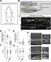
BYN-1 is identified as a binding partner of GLH-1. (A) Schematic of bystin/BYN-1 proteins in C. elegans and Homo sapiens, with the bystin coiled-coil domain indicated in black. (B) Schematic of the conserved FG domain in C. elegans GLH-1 and H. sapiens trophinin. FG, phenylalanine-glycine repeats (black dots represent FG-repeats); MAGE, melanoma antigen gene domain. (C) Directed 1×1 Y2H assay shows that the C terminus of BYN-1 binds full-length GLH-1, but the FG-repeat domain of GLH-1 is not sufficient to bind BYN-1. Removal of the FG-repeat domain from GLH-1 weakens binding to BYN-1 when stringency is challenged with increasing concentrations of 3-aminotriazole (3AT).
Fig. 2.
Fig. 2.
BYN-1 affects cell polarity and migration and is essential for fertility and development. (A) Brood size comparison of wild-type N2 L1 worms fed control RNAi (L4440; ctrl) or RNAi targeting byn-1 (Vidal byn-1 RNAi clone, pDU103 byn-1 N-terminal RNAi clone). Number of analyzed animals: n=14 for N2 (ctrl), n=14 for N2 (byn-1 RNAi-Vidal), n=12 for N2 (ctrl), n=14 for N2 (byn-1 RNAi-pDU103). (B) Brood size comparison of wild-type N2 L1 and L4 worms fed control RNAi (L4440) or byn-1 RNAi (Vidal). Number of analyzed animals: n=12 for the respective conditions. (C) Embryonic lethality from wild-type N2 L4 worms fed control RNAi (L4440) or byn-1 RNAi (Vidal). Number of analyzed mothers: n=12 for N2 (ctrl), n=11 for N2 (byn-1 RNAi). (D) DIC images of embryos from the dissected wild-type adults that had been fed control (L4440) or byn-1 RNAi (Vidal) at L4 stage. Embryos were classified into four categories: (1) ovoid, (2) circular, (3) ruptured or (4) small. Scale bars: 20 µm. (E) The eggshells of progeny from L4 animals fed byn-1 RNAi, but not control RNAi, are permeable to the lipophilic dye FM1-43 (arrows). Scale bar: 20 µm. n=number of embryos. (F) Depletion of byn-1 in L4 animals causes abnormal GLH-1::GFP localization in live embryos. Scale bar: 10 µm. Graph shows the percentage of two-cell embryos with loss of GLH-1 polarity caused by depletion of byn-1. Number of analyzed two-cell embryos: n=34 for ctrl, n=38 for byn-1 RNAi. (G) The developmental stage of the F1 generation from L4 stage worms fed control or byn-1 RNAi every 24 h at 20°C. Ad, adult (with embryos); YA, young adult (with no embryos). Number of analyzed animals: n=60 for N2 (ctrl), n=62 for N2 (byn-1 RNAi), n=50 for rrf-3 (ctrl), n=66 for rrf-3 (byn-1 RNAi). (H) F1 progeny from CRISPR-generated glh-1::GFP I; Δbyn-1 III hermaphrodites balanced over hT2 g(qIs48-green pharynx) I/III, 3 days post-egg-lay. Δbyn-1 homozygous animals (no green pharynx) are outlined in magenta and show fully penetrant primordial germ cell (glh-1::GFP) and L1 arrest phenotypes. In A-C,F, error bars represent s.d. ***P<0.001 (two-tailed, unpaired Student's t-test).
Fig. 3.
Fig. 3.
BYN-1 and GLH-1 localization in germ cells. (A) Top: Schematic of a hermaphrodite gonad arm in C. elegans adults. In the gonad arm, germ cells undergo mitotic proliferation in the distal ‘mitotic zone’ (MZ). During their passage through the ‘transition zone’ (TZ), germ cells stop dividing and initiate meiosis. Germ cells in meiotic prophase I accumulate in the ‘pachytene zone’ (PZ). The ‘loop region’ is the anatomic bend in the gonad. The more proximal region of the gonad arm is filled with developing oocytes. Bottom: Confocal live images showing BYN-1::mCherry and GLH-1::GFP during oogenesis in adult hermaphrodites (20°C). GLH-1 is located on the cytoplasmic surface of the nuclear periphery, and BYN-1 is in the nucleolus during oogenesis. Scale bar: 5 µm. (B) Top: Schematic of a male gonad arm in C. elegans adults. Following an extended pachytene stage, spermatocytes enter the karyosome stage in the condensation zone (CZ) before detaching from the syncytial germline. Meiotic divisions in the division zone (DZ) are followed by spermatids budding from anucleate residual bodies (RBs) that become engulfed and cleared by the somatic gonad surrounding the seminal vesicle. Bottom: Confocal live images showing the dynamic localization of BYN-1::mCherry and GLH-1::GFP in germ cells from adult males (20°C). BYN-1 is nucleolar from MZ to PZ but translocates into the cytoplasm at CZ to colocalize with GLH-1. The MZ, TZ and PZ panels represent a single confocal plane, whereas the CZ panels represent a maximum-intensity z-projection to capture the change in BYN-1 dynamics. Scale bars: 5 µm. (C) Enlarged confocal live images of GLH-1 and BYN-1 in respective germ cell stages of male germline. Scale bar: 3 µm. (D) Enlarged confocal live images of GLH-1 and BYN-1 in respective germ cell stages of hermaphrodite germline. Scale bar: 3 µm. (E) Representative live images of GLH-1 and BYN-1 in dissected male germ cells during the meiotic and post-meiotic events of spermatogenesis. The graph shows the percentage of colocalization of GLH-1 and BYN-1 among cells in respective stages of spermatogenesis (n=30 per stage). Error bars represent s.d. AI/AII, anaphase I/II; D, diplotene; MI/MII, metaphase I/II; n.d., no data.
Fig. 4.
Fig. 4.
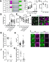
BYN-1 contributes to the clearance of RBs during spermatogenesis. (A) DAPI-stained adult male dissected gonads labeled by developmental stages. Scale bar: 10 µm. (B) GLH-1::GFP expression in control (ctrl) RNAi and byn-1 RNAi-treated adult stage males. Male gonads were fixed and counterstained with DAPI (blue). Scale bar: 10 µm. d, distal region of the gonad. Graph shows the number of GLH-1 containing RBs in each male gonad. n=14 for ctrl, n=15 for byn-1 RNAi. (C) Dissected gonads from wild-type control and byn-1 RNAi males co-stained with anti-α-tubulin (green), anti-phosphorylated histone H3 Ser10 (magenta), and DAPI (blue). Scale bar: 10 µm. d, distal region of the gonad. Graph shows the number of α-tubulin-positive RBs at the proximal region of gonad. n=16 for ctrl, n=17 for byn-1 RNAi. (D) Representative live images of dissected sperm showing that RBs stain with Annexin V (green). Graph shows the number of Annexin V-positive RBs. n=15 for ctrl, n=15 for byn-1 RNAi. Scale bar: 10 µm. (E) Sperm numbers were quantified in males of each group. n=9 for ctrl, n=11 for byn-1 RNAi. (F) Cross progeny counts from fog-2 females mated with control RNAi and byn-1 RNAi males. n=10 for ctrl, n=10 for byn-1 RNAi. (G,H) RB (α-tubulin positive) counts in the proximal region of dissected male gonads. n=25 for the respective conditions. Error bars represent s.d. Statistical significance was calculated using a two-tailed, unpaired Student's t-test ***P<0.001.
Fig. 5.
Fig. 5.
BYN-1 functions upstream of CED-1 during RB clearance. (A) Engulfment pathways for RB clearance during spermatogenesis as described by Huang et al. (2012). PS, phosphatidylserine. (B) Schematic of a gonad arm in C. elegans L4-larval-stage to adult hermaphrodite. In the L4 stage, RBs at the proximal region of the gonad are surrounded by CED-1 (green circle with white border), a transmembrane receptor that mediates cell corpse engulfment and clearance of RBs during spermatogenesis. In adults, apoptotic germ cells are mostly observed in the late pachytene zone surrounded by CED-1 (black circle with yellow border) during oogenesis. (C) RB engulfment visualized with smIs34[ced-1p::ced-1::GFP] transgene at 20°C. Engulfed RBs surrounded by CED-1::GFP are marked with white arrows. Yellow arrows mark the engulfment of germ cell corpses. byn-1 RNAi adult animals showed both engulfed RBs at the proximal region of the gonad and engulfed germ cell corpses at the pachytene zone. d, distal region of gonad. Scale bar: 20 µm. (D,E) The number of RBs surrounded by CED-1::GFP per gonad arm at 6 h and 24 h post-L4 stage at 20°C. ctrl, control. n=18-20 for the respective conditions. (F,G) The number of RBs, as determined by DIC microscopy (H), in either ced-1(e1735) or ced-12 (n3261) hermaphrodites 24 h post-L4 stage. n=16-22 for the respective conditions. (H) DIC images of RB retention in the spermatheca of 24 h post-L4 stage ced-1 and ced-12 hermaphrodites. Yellow dashed boxes indicate the approximate region that is enlarged to the right. Arrowheads represent RBs. Scale bar: 10 µm. Error bars represent s.d. Statistical significance was calculated using a two-tailed, unpaired Student's t-test. ***P<0.001. n.s., not significant (P>0.05).
Fig. 6.
Fig. 6.
The FG-repeat domains of GLH-1 and GLH-2 contribute to RB clearance. (A) Schematic of endogenous GLH-1::GFP and GLH-2 modified proteins in wild-type (WT), ΔFG and 2ΔFG background strains. (B) GFP-expressing RBs were scored 6 h or 24 h post-L4 stage in hermaphrodites. n=15-23 for the respective strains. (C) Sperm number in WT, ΔFG and 2ΔFG young adult (YA) hermaphrodites. n=10-11 for the respective strains. (D) RBs were scored in respective genetic backgrounds with control and byn-1 RNAi at 24 h in post-L4 stage hermaphrodites. n=31-40 for the respective conditions. (E) The number of sperm was quantified in respective genetic backgrounds with control and byn-1 RNAi in YA hermaphrodites. n=11-19 for the respective conditions. (F) Confocal live images showing the localization of GLH-1::GFP and BYN-1::mCherry at the condensation zone in WT and 2ΔFG mutant males. Left: Male spermatogenesis at 20°C. Scale bar: 5 μm. Right: Enlarged diplotene and karyosome stages of spermatogenesis in WT and 2ΔFG mutants. Scale bar: 3 µm. (G) Colocalization of GLH-1::GFP and BYN-1::mCherry decreases in the condensation zone of 2ΔFG mutants. n=15 for each strain. (H) GLH-1::GFP-granule sizes at the condensation zone in the male germline quantified using ImageJ. n=40 for each strain. (I) Enlarged confocal live images of GLH-1::GFP and BYN-1::mCherry in the RBs of WT and 2ΔFG mutants. Scale bar: 3 µm. (J) Quantification of BYN-1::mCherry intensity in RBs of WT and 2ΔFG mutant males. n=9 for each strain. (K) Colocalization of GLH-1::GFP and BYN-1::mCherry in RBs of WT and 2ΔFG mutants. n=11 for each strain. Statistical significance for B-E calculated using one-way ANOVA, and for G,H,J,K a two-tailed, unpaired Student's t-test. ***P<0.001, *P<0.05. Error bars represent s.d.
Fig. 7.
Fig. 7.
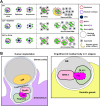
Working model for BYN-1/bystin-mediated cellular engulfment. (A) The cytoplasmic translocation of BYN-1 appears to be required for activation of CED-1 for residual body (RB) engulfment during spermatogenesis. Upon nucleolar breakdown in the diplotene stage of spermatogenesis, BYN-1 disperses throughout the nucleoplasm. In the karyosome stage, BYN-1 leaves the nucleus where it colocalizes with GLH-1 in perinuclear germ granules. During the budding stage of spermatogenesis, GLH-1 and BYN-1 are deposited in anucleate RBs that become engulfed by CED-1-positive cells of the overlying somatic gonad. WT, wild type. (B) Similarities between human embryonic implantation and C. elegans RB clearance point to possible conservation of pathways that regulate cellular uptake. ICM, inner cell mass.

References

    1. Adachi, K., Soeta-Saneyoshi, C., Sagara, H. and Iwakura, Y. (2007). Crucial role of Bysl in mammalian preimplantation development as an integral factor for 40S ribosome biogenesis. Mol. Cell. Biol. 27, 2202-2214. 10.1128/MCB.01908-06 - DOI - PMC - PubMed
    1. Aoki, R., Suzuki, N., Paria, B. C., Sugihara, K., Akama, T. O., Raab, G., Miyoshi, M., Nadano, D. and Fukuda, M. N. (2006). The Bysl gene product, bystin, is essential for survival of mouse embryos. FEBS Lett. 580, 6062-6068. 10.1016/j.febslet.2006.09.072 - DOI - PMC - PubMed
    1. Aoyama, J., Nakayama, Y., Sugiyama, D., Saburi, S., Nadano, D., Fukuda, M. N. and Yamaguchi, N. (2005). Apical cell adhesion molecule, trophinin, localizes to the nuclear envelope. FEBS Lett. 579, 6326-6332. 10.1016/j.febslet.2005.10.012 - DOI - PubMed
    1. Arai, Y., Hosoda, F., Kobayashi, H., Arai, K., Hayashi, Y., Kamada, N., Kaneko, Y. and Ohki, M. (1997). The inv(11)(p15q22) chromosome translocation of de novo and therapy-related myeloid malignancies results in fusion of the nucleoporin gene, NUP98, with the putative RNA helicase gene, DDX10. Blood 89, 3936-3944. 10.1182/blood.V89.11.3936 - DOI - PubMed
    1. Ayala, G. E., Dai, H., Li, R., Ittmann, M., Thompson, T. C., Rowley, D. and Wheeler, T. M. (2006). Bystin in perineural invasion of prostate cancer. Prostate 66, 266-272. 10.1002/pros.20323 - DOI - PubMed

MeSH terms

Substances

LinkOut - more resources