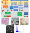
Asbestos exposure diagnosis in pulmonary tissues
- PMID: 39377502
- PMCID: PMC11460150
- DOI: 10.32074/1591-951X-930
Asbestos exposure diagnosis in pulmonary tissues
Abstract
The diagnosis of asbestosis requires different criteria depending on whether it is in a clinical or medical/legal setting. In the latter context, only when a "diffuse interstitial fibrosis associated to asbestos bodies (ABs)" is present, it can be said to be asbestosis. Considering the medical/legal setting, the diagnosis must be certain and proven. Unfortunately, it is often difficult to identify ABs by light microscopy (LM), but this does not mean that the diagnosis should be clinically excluded. Other parameters are important, such as working history and/or diagnostic imaging. In addition to LM, normally used for diagnosis, there are other techniques, e.g.: scanning electron microscopy with attached microanalysis microprobe (SEM/EDS), but they require tissue digestion and higher cost. A new approach with micro-Raman spectroscopy and SEM/EDS techniques is able to analyse histological sections without other manipulations that could interfere with analysis of asbestos fibres. In this work, we propose an algorithm for asbestosis diagnosis, especially in the forensic medical field, demonstrating the importance of close collaboration between multiple professionals.
Keywords: asbestos; diagnosis; lung; mineralogist; pathologist.
Copyright © 2024 Società Italiana di Anatomia Patologica e Citopatologia Diagnostica, Divisione Italiana della International Academy of Pathology.
Conflict of interest statement
The authors declare no conflict of interest.
Figures

References
-
- Sporn TA, Roggli VL. Asbestosis. In: Oury TD, Sporn TA, Roggli VL. (Ed.) Pathology of Asbestos-Associated Diseases. Third Edition. Springer-Verlag Berlin Heidelberg, 2014, pp. 53-80. https://doi.org/10.1289/ehp.80341 10.1289/ehp.80341 - DOI
-
- Cooke WE. Fibrosis of the lungs due to the inhalation of asbestos dust. Br Med J 1924;2(3317):147-148. https://doi.org/10.1136/bmj.2.3317.147 10.1136/bmj.2.3317.147 - DOI - PMC - PubMed
-
- Grosso F, Croce A, Trincheri NF, et al. . Asbestos fibres detected by scanning electron microscopy in the gallbladder of patients with malignant pleural mesothelioma (MPM). J Microsc 2017;266:48-54. https://doi.org/10.1111/jmi.12517 10.1111/jmi.12517 - DOI - PubMed
-
- Croce A, Capella S, Belluso E, et al. . Asbestos fibre burden in gallbladder: a case study. Micron 2018;105:98-104. https://doi.org/10.1016/j.micron.2017.12.001 10.1016/j.micron.2017.12.001 - DOI - PubMed
-
- Grosso F, Croce A, Libener R, et al. . Asbestos fiber identification in liver from cholangiocarcinoma patients living in an asbestos polluted area: a preliminary study. Tumori J 2019;105:404-410. https://doi.org/10.1177/0300891619839305 10.1177/0300891619839305 - DOI - PubMed
Publication types
MeSH terms
Substances
LinkOut - more resources
Full Text Sources
Medical

