. 2024 Oct 1;13(10):14.
doi: 10.1167/tvst.13.10.14.

IOP Reduction in Nonhuman Primates by Microneedle Injection of Drug-Free Hydrogel to Expand the Suprachoroidal Space

Affiliations

IOP Reduction in Nonhuman Primates by Microneedle Injection of Drug-Free Hydrogel to Expand the Suprachoroidal Space

Yooree G Chung et al. Transl Vis Sci Technol. .

Abstract

Purpose: Expansion of the suprachoroidal space (SCS) by a hydrogel injection has been shown to reduce intraocular pressure (IOP) in rabbits as a potential treatment for ocular hypertension in glaucoma. Here, we evaluate the safety and efficacy of this approach in hypertensive and normotensive eyes in nonhuman primates.

Methods: A microneedle was used to inject a hyaluronic acid-based hydrogel or saline solution (control) into the SCS of cynomolgus monkey eyes that were either normotensive (n = 7 experimental; n = 2 control eyes) or had induced ocular hypertension (n = 6 experimental; n = 3 control eyes). IOP and the degree of SCS expansion were monitored over time by tonometry and ultrasound biomicroscopy, respectively. Safety was evaluated through slit lamp, fundus, and histology examinations.

Results: In hypertensive eyes, SCS injection with hydrogel initially reduced IOP by 47.5 ± 16.7%, and IOP returned to baseline in 38 days. In normotensive eyes, hydrogel injection initially reduced IOP by 38.8 ± 8.1% and IOP gradually returned to baseline also in 39 days. Sham injections resulted in mild IOP reduction in hypertensive eyes and normotensive eyes. The hydrogel injections were well tolerated by clinical assessments.

Conclusions: IOP was reduced in nonhuman primates for over one month by sustained SCS expansion. This procedure was safe and simple to perform. These data confirm the translational potential of this treatment method. Further optimization of the hydrogel may provide longer durations of IOP reduction.

Translational relevance: A microneedle injection of hydrogel into the suprachoroidal space may provide a non-surgical, non-pharmacologic treatment for ocular hypertension in glaucoma patients.


Conflict of interest statement

Disclosure: Y.G. Chung, None; S. Fan, None; V. Gulati, None; H.-L. Li, None; H. Gong, None; C.B. Toris, None; M.R. Prausnitz, Clearside Biomedical, Inc. (I, O), US20230201110A1 (P); C.R. Ethier, Equinox Ophthalmic (C), Injectsense (I), US20230201110A1 (P)

Figures

Figure 1.
Figure 1.
Representative images of a microneedle and hydrogel used for injections. (A) Microneedle (arrow) connected to a 1 mL syringe. (B) Microneedles were 900 to 1000 µm in length. (C) A representative volume of 50 µL hydrogel after gelation in vitro.
Figure 2.
Figure 2.
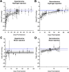
Effect of hydrogel injection on intraocular pressure and SCS expansion. Hypertensive and normotensive monkey eyes were injected with the crosslinked hydrogel (A and B, respectively) or a sham injection of Hanks’ Balanced Salt Solution (C and D, respectively). Change in IOP compared to pre-injection baseline values (Delta IOP) is plotted. Delta baseline IOP means (blue solid lines, equal to zero by definition) and confidence intervals (blue dotted lines; see text) are shown in each panel. In panels A, B, and D, the regression fit (solid black line) and confidence band (gray area) are shown; the intersection of the confidence band and the mean Delta baseline IOP is indicated by the black box, which indicates the time point at which the Delta IOP was no longer significantly different from zero. In panel C, the mean Delta IOP (solid black line) and confidence intervals (gray area) are shown in black. Plotted data are the average ± SD of six (A, hydrogel), three (C, sham), seven (B, hydrogel) or two (D, sham) replicates.
Figure 3.
Figure 3.
Representative ultrasound biomicroscopy images of the SCS after hydrogel injection. (A) Hypertensive and (B) normotensive eyes are shown after injection with crosslinked hydrogel or sham injection of Hanks’ Balanced Salt Solution. All images are in the same orientation (C, cornea; I, iris; CB, ciliary body; Ch, choroid; H, hydrogel; S, sclera). The dotted yellow line approximately outlines the hydrogel in the SCS; these images were used to determine EWT. Each injection type (sham or hydrogel) across time points is from the same eye. Images are representative of three sham eyes and six hydrogel eyes for hypertensive animals and two sham eyes and seven hydrogel eyes for normotensive animals.
Figure 4.
Figure 4.
Correlation of Delta IOP versus Delta EWT. Delta IOP (circles; right axis) with linear regression fit and confidence band (black line and gray area; right axis) and Delta EWT (square; left axis) are shown for (A) hypertensive and (B) normotensive eyes. A cross-plot of Delta EWT versus Delta IOP (data from graphs A and B) are shown for (C) hypertensive and (D) normotensive hydrogel-injected eyes. Both hypertensive (linear regression R2 = 0.26, Pearson r = −0.51, P = 0.0005) and normotensive (R2 = 0.60, Pearson r = −0.78, P < 0.0001) eyes demonstrated that Delta IOP was negatively correlated with Delta EWT. In panels C and D, the solid lines represent the linear regression fits, with the gray-shaded areas representing the 95% confidence bands of the linear regressions.
Figure 5.
Figure 5.
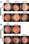
Representative fundoscopic images of eyes after hydrogel injection. (A) Hypertensive and (B) normotensive eyes are shown after injection with crosslinked hydrogel or sham injection of Hanks’ balanced salt solution. Images are from the same eye across time points for each injection type. Images are representative of three sham eyes and six hydrogel eyes for hypertensive animals and two sham eyes and seven hydrogel eyes for normotensive animals. No changes in retinal morphology due to injection were identified at any timepoint. N, nasal; T, temporal.
Figure 6.
Figure 6.
Representative photographs of the external eye after hydrogel injection. (A) The injected eye immediately before and after injection and (B) both injected and control eyes on day 1 and day 2 after injection. Hydrogel was injected in the inferotemporal quadrant of the left eye, and the injection site was visible immediately after injection (arrow). Mild edema occurred one day after injection and was nearly resolved by the second day. N, nasal; S, superior.
Figure 7.
Figure 7.
Low- and medium-magnification representative histologic images of ocular tissue collected at least three months after SCS injection. (A, B) A noninjected ocular hypertensive eye (negative control). (C, D) A saline-injected normotensive eye at the injection area. (E, F) A hydrogel-injected normotensive eye at the injection area. Images A, C, and E were captured under magnification ×4, whereas images B, D, and F show further magnification of the boxed areas 3 mm from the limbus (approximate location of injection) captured under magnification ×20. Expansion of the SCS is likely an artefact of histology preparation, with no evidence of active fibrosis or inflammation at the injection site. Variations in pigment could be observed but were within the normal range in monkeys.
Figure 8.
Figure 8.
High-magnification representative histologic images of ocular tissue after injection. (A, B) A normotensive eye injected with 100 µL hydrogel. Images are at the injection area three months after injection. (C, D) A hypertensive eye injected with 50 µL of hydrogel. Images are at the injection area eight months after injection. Images A and C were captured under magnification ×4, whereas images B and D show magnification ×40 of the boxed areas 3 mm from the limbus (approximate location of injection). Blue arrows indicate gel residue, and the yellow arrow points to the giant cells.

References

    1. Weinreb RN, Aung T, Medeiros FA.. The pathophysiology and treatment of glaucoma: a review. JAMA. 2014; 311: 1901–1911. - PMC - PubMed
    1. Quigley HA, Broman AT.. The number of people with glaucoma worldwide in 2010 and 2020. Br J Ophthalmol. 2006; 90: 262–267. - PMC - PubMed
    1. Tham YC, Li X, Wong TY, Quigley HA, Aung T, Cheng CY.. Global prevalence of glaucoma and projections of glaucoma burden through 2040: a systematic review and meta-analysis. Ophthalmology. 2014; 121: 2081–2090. - PubMed
    1. Collaborative Normal-Tension Glaucoma Study Group. The effectiveness of intraocular pressure reduction in the treatment of normal-tension glaucoma. Am J Ophthalmol. 1998; 126: 498–505. - PubMed
    1. Kass MA, Heuer DK, Higginbotham EJ, et al. .. The Ocular Hypertension Treatment Study: a randomized trial determines that topical ocular hypotensive medication delays or prevents the onset of primary open-angle glaucoma. Arch Ophthalmol (Chicago, Ill 1960). 2002; 120: 701–713; discussion 829–830. - PubMed

Publication types

LinkOut - more resources