. 2024 Dec;60(6):696-710.
doi: 10.1007/s11262-024-02112-4. Epub 2024 Oct 8.

Identification and characterization of mycoviruses in transcriptomes from the fungal family ceratocystidaceae

Affiliations

Identification and characterization of mycoviruses in transcriptomes from the fungal family ceratocystidaceae

Bianca Hough et al. Virus Genes. 2024 Dec.

Abstract

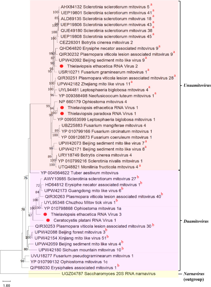
Mycoviruses pervade the fungal kingdom, yet their diversity within various fungal families and genera remains largely unexplored. In this study, 10 publicly available fungal transcriptomes from Ceratocystidaceae were analyzed for the presence of mycoviruses. Despite mycovirus associations being known in only four members of this family, our investigation unveiled the discovery of six novel mycoviruses. The majority of these mycoviruses are composed of positive sense single stranded RNA and are putatively assigned to the viral family Mitoviridae (with tentative classification into the genera Unuamitovirus and Duamitovirus). The double stranded RNA viruses, however, were associated with the family Totiviridae (with tentative classification into the genus Victorivirus). This study also revealed the discovery of an identical unuamitovirus in the fungal species Thielaviopsis ethacetica and Thielaviopsis paradoxa. This discovery was notable as these fungal isolates originated from distinct geographical locations, highlighting potential implications for the transmission of this mitovirus. Moreover, this investigation significantly expands the known host range for mycoviruses in this family, marking the initial identification of mycoviruses within Ceratocystis platani, Thielaviopsis paradoxa, Thielaviopsis ethacetica, and Huntiella omanensis. Future research should focus on determining the effects that these mycoviruses might have on their fungal hosts.

Keywords: Ceratocystis; Thielaviopsis; Ceratocystidaceae; Mitoviruses; Mycoviruses.


Conflict of interest statement

Declarations Conflict of interest The authors declare no competing interests. Ethical approval The manuscripts constitutes original research and has not been previously published. All authors are in agreement with manuscript submission, which complies with the ethical standards of the journal.

Figures

Fig. 1
Fig. 1
Phylogenetic tree showing maximum likelihood analysis of the RdRp protein sequences from the mitoviruses discovered in this study, with those of related viruses. The phylogeny was generated using IQ-TREE, using the Whelan and Goldman model with frequencies, invariant sites, and gamma distribution. Bootstrapping with 1000 replicates was performed, and corresponding percentages are indicated next to the branches. Only bootstrap values equal to or exceeding 70 are displayed. The Saccharomyces 20S RNA Narnavirus was used as an outgroup. The red circles (●) indicate mycoviruses discovered in this study. a Denotes unuamitoviruses and b: duamitoviruses which have not yet been taxonomically assigned by the ICTV (https://ictv.global/taxonomy). These mitoviruses exhibit genetic characteristics and phylogenetic relationships that strongly suggest their classification within these genera
Fig. 2
Fig. 2
Percent Identity Matrix of the mitoviruses from this study, and other viral members from Mitoviridae, generated using Clustal Omega. The color gradients represent percent identities, with darker blue shades indicating higher identities and lighter shades indicating lower identities. The mitoviruses are numbered from 1 to 13, and their corresponding labels are displayed at the bottom of each column. Mitoviruses from this study are highlighted in red for better readability
Fig. 3
Fig. 3
Schematic representation of the genomic organization of a TpRV-1, b TeRV-1, c TeRV-2, d TeRV-3, and e CpRV-1. The open bars represent single open reading frames (ORFs), while the single lines represent untranslated regions (UTRs). The length of the ORF is indicated for all mitoviruses, and the position of the RdRp domain encoded on the ORF is also shown
Fig. 4
Fig. 4
Maximum likelihood phylogenetic tree constructed from the alignment of RdRp protein sequences from totiviruses discovered in this study, with those of related viruses. The phylogeny was generated using IQ-TREE, using the Le Gauss model with frequencies, invariant sites, and gamma distribution. Bootstrapping with 1000 replicates was performed, and corresponding percentages are indicated next to the branches. Only bootstrap values equal to or exceeding 70 are displayed. The Saccharomyces cerevisiae virus L-A was used as an outgroup. The red circles (●) indicate the totiviruses discovered in this study
Fig. 5
Fig. 5
Percent Identity Matrix of the totiviruses from this study, and other viral members from the genus Victorivirus. The Matrix was generated using Clustal-Omega 1.2.2, with the color gradients serving to represent the respective percent identities of the totiviruses from this study to other related viruses. The percent identity matrixes are presented as A, denoting the alignments of the a RdRp and b those of the CP amino acid sequences. The percent identities are visualized with varying shades of blue and pink in the RdRp and CP alignments, respectively. Higher percentages are indicated in darker shades, while lower percentages are indicated in lighter hues. For clarity, the totiviruses from this study are numbered as 1–7 in the CP alignments and as 1–8 in the RdRp alignments. Corresponding labels for each of these totiviruses
Fig. 6
Fig. 6
Schematic representation of the genome organization of TpRV-2. The open bars signify individual ORFs, and the single lines represent the 5’ and 3’ UTRs. The putative victorivirus consists of two ORFs, and their respective lengths are indicated. Moreover, the positions of the RdRp and CP domains encoded within their corresponding ORFs are shown. The reading frames for each ORF are clearly indicated, and the stop-initiation codon required for the translation of ORF 2 is also depicted in the figure

Similar articles

References

    1. Fenner FJ, McAuslan BR, Mims C (2013) The biology of animal viruses. Elsevier, Amsterdam
    1. Hull R (2013) Plant virology. Academic Press, New York
    1. Weinbauer MG (2004) Ecology of prokaryotic viruses. FEMS Microbiol Rev 28:127–181. 10.1016/j.femsre.2003.08.001 - PubMed
    1. La Scola B (2014) Looking at protists as a source of pathogenic viruses. Microb Pathog 77:131–135. 10.1016/j.micpath.2014.09.005 - PubMed
    1. Ghabrial SA, Suzuki N (2009) Viruses of plant pathogenic fungi. Annu Rev Phytopathol 47:353–384 - PubMed

LinkOut - more resources