. 2024 Oct 15;121(42):e2309548121.
doi: 10.1073/pnas.2309548121. Epub 2024 Oct 8.

YTHDC2 serves a distinct late role in spermatocytes during germ cell differentiation

Affiliations

YTHDC2 serves a distinct late role in spermatocytes during germ cell differentiation

Alexis S Bailey et al. Proc Natl Acad Sci U S A. .

Abstract

Posttranscriptional regulation of gene expression by RNA-binding proteins can enhance the speed and robustness of cell state transitions by controlling RNA stability, localization, or if, when, or where mRNAs are translated. The RNA helicase YTHDC2 is required to shut down components of the mitotic program to facilitate a proper switch from mitosis to meiosis in mouse germ cells. Here, we show that YTHDC2 has a second essential role in promoting meiotic progression in late spermatocytes. Inducing conditional knockout of Ythdc2 during the first wave of spermatogenesis, after initiation of meiotic prophase, allowed YTHDC2-deficient germ cells to advance to the pachytene stage and properly express many meiotic markers. However, the YTHDC2-deficient spermatocytes mis-expressed a number of genes, some up-regulated and some down-regulated, failed to transition to the diplotene stage, and then quickly died. Coimmunoprecipitation experiments revealed that YTHDC2 interacts with several RNA-binding proteins in early or late spermatocytes, with many of the interacting proteins, including MEIOC, localizing to granules, similar to YTHDC2. Our findings suggest that YTHDC2 collaborates with other RNA granule components to facilitate proper progression of germ cells through multiple steps of meiosis via mechanisms influencing posttranscriptional regulation of RNAs.

Keywords: RNA-binding proteins; YTHDC2; germ cells; meiosis; spermatocytes.


Conflict of interest statement

Competing interests statement:The authors declare no competing interest.

Figures
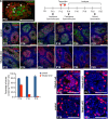
Fig. 1.
Fig. 1.
Knock out of Ythdc2 in spermatocytes during the first wave of spermatogenesis. (A) Immunofluorescence image of a P18 wild-type mouse testis tubule cross-section stained for YTHDC2 (green) and SYCP3 (red). Arrowheads: leptotene spermatocytes from the second wave of spermatogenesis. Arrows: pachytene spermatocytes from the first wave of spermatogenesis within the lumen of a testis tubule. (B) Timeline of initial appearance of germ cell stages during the first wave of spermatogenesis, with the timing of tamoxifen injections given to both control and Ythdc2 cKO mice. (C-N) Immunofluorescence images of control (+/+; Ythdc2flox/Δ, Top) and Ythdc2 cKO (UBC-CreERT2; Ythdc2flox/Δ, Bottom) seminiferous tubule cross-sections from (C and D) P12, (E and F) P14, (G and H) P16, (I and J) P18, (K and L) P20, and (M and N) P25 testes stained with anti-YTHDC2 (green) and anti-SYCP3 (red) antibodies as well as DAPI to mark the DNA (blue). (O) Percentage of tubules containing YTHDC2-positive cells in P14, P16, and P18 control (+/+; Ythdc2flox/Δ) (blue) and Ythdc2 cKO (red) testes (n = 2 to 4 mice per group; t test, P14: P-value = 0.0003, P16: P-value < 0.0001, P18: P-value = 0.0019). Error bars: SEM. (PS) Immunofluorescence images of testis tubules from (P) P12 Ythdc2 null, (Q) P12 Ythdc2 cKO, (R) P16 control (+/+; Ythdc2flox/Δ) and (S) P16 Ythdc2 cKO mice stained with anti-CYCLINA2 (red) antibody and DAPI (blue). (Scale bars, 50 µm.)
Fig. 2.
Fig. 2.
YTHDC2 is required for late spermatocytes to progress through meiotic prophase. (AL) Sections of mouse seminiferous tubules stained with periodic acid–Schiff (PAS) from (A and B) P12, (C and D) P14, (E and F) P16, (G and H) P18, (I and J) P20, and (K and L) P25 tamoxifen-treated control (+/+; Ythdc2flox/Δ, Top) and Ythdc2 cKO (UBC-CreERT2; Ythdc2flox/Δ, Bottom) testis. (Scale bars, 50 µm.) (MP) Testis from P16 control (+/+; Ythdc2flox/Δ) and Ythdc2 cKO (UBC-CreERT2; Ythdc2flox/Δ) mice stained with (M and N) anti-YTHDC2 (green), anti-SYCP3 (red) antibodies as well as DAPI to mark the DNA (blue) or (O and P) anti-YTHDC2 (green), anti-γH2AX (red) antibodies and DAPI (blue). (Q) Percentage of tubule cross-sections containing spermatogonia (Spg), preleptotene (Pre-L), leptotene (Lep), zygotene (Zyg), pachytene (Pach), or diplotene (Dip) as the most advanced cell stage in P16 and P18 control (+/+; Ythdc2flox/Δ) and Ythdc2 cKO (UBC-CreERT2; Ythdc2flox/Δ) testes (n = 2 mice per group; >120 tubules counted per mouse). Error bars: SEM. (RW) Testis tubule cross-sections from (RT) P16 and (UW) P18 Cre control (UBC-CreERT2; Ythdc2flox/+), flox/Δ control (+/+; Ythdc2flox/Δ), and Ythdc2 cKO (UBC-CreERT2; Ythdc2flox/Δ) mice labeled with TUNEL (red) and VASA (green). Arrow: Tubule filled with TUNEL-positive cells. (Scale bars, 50 µm.) (X) Number of TUNEL-positive cells per tubule cross-section in P14, P16, and P18 Cre control (light blue), flox/Δ control (dark blue), and Ythdc2 cKO (red) testes (n = 2 to 4 mice per group; >110 tubules counted per mouse; t test, *P-value = 0.0176, **P-value = 0.0268). Error bars: SEM.
Fig. 3.
Fig. 3.
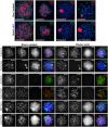
Spermatocytes lacking YTHDC2 reach the late pachytene stage but then quickly die. (AH) Immunofluorescence images of germ cell spreads from P18 tamoxifen-treated control (+/+; Ythdc2flox/Δ, Top) and Ythdc2 cKO (UBC-CreERT2; Ythdc2flox/Δ, Bottom) mice stained with anti-SYCP3 (green), anti-γH2AX (red) antibodies and DAPI to mark the DNA (blue). (IL‴) Immunofluorescence images of germ cell spreads from P16 control (+/+; Ythdc2flox/Δ, Left) and Ythdc2 cKO (UBC-CreERT2; Ythdc2flox/Δ, Right) mice stained with anti-SYCP3 (green), anti-HORMAD1 (red) antibodies and DAPI (blue). Arrows: HORMAD1 staining on the sex body in pachytene spermatocytes. (MT‴) Germ cell spreads from P16 control (+/+; Ythdc2flox/Δ, Left) and Ythdc2 cKO (UBC-CreERT2; Ythdc2flox/Δ, Right) mice stained with anti-SYCP3 (green), anti-SYCP1 (red), and anti-H1t (blue) antibodies.
Fig. 4.
Fig. 4.
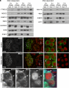
YTHDC2 interacts with several other proteins involved in RNA regulation. (A and B) IP with α-YTHDC2 from P12 and P18 wild-type mouse testis extracts treated either with RNase Inhibitor (−RNase) or RNase A (+RNase). Western blots probed with anti-YTHDC2, anti-MEIOC, anti-PABPC1, anti-FMR1, anti-CAPRIN1, anti-EWS, anti-PTBP1, anti-MOV10, anti-YBX1, anti-G3BP2, and anti-UPF1 antibodies. (CH) Immunofluorescence images of testis cross-sections from P18 (CE) control (+/+; Ythdc2flox/Δ) and (FH) Ythdc2 cKO (UBC-CreERT2; Ythdc2flox/Δ) mice stained with anti-MEIOC (green) and anti-SYCP3 (red) antibodies. (Scale bars, 50 µm.) (D and E) High-magnification images of (D) pachytene spermatocytes (first wave) in boxed region 1 in panel C′ and (E) leptotene spermatocytes (second wave) in boxed region 2 in panel C′. (G and H) High-magnification images of (G) pachytene spermatocytes (first wave) in boxed region 1 in panel F′ and (H) leptotene spermatocytes (second wave) in boxed region 2 in panel F′. Arrows: MEIOC protein in perinuclear puncta in pachytene spermatocytes. (II‴) High-magnification immunofluorescence images of spermatocytes from a P18 Ythdc2 cKO mouse testis cross-section stained with anti-MEIOC (green), anti-MIWI (blue), and anti-DCP1A (red) antibodies. Arrows: puncta in the cytoplasm of pachytene spermatocytes. (Scale bar, 5 µm.)
Fig. 5.
Fig. 5.
Early transcript changes following conditional knockout of Ythdc2 in first wave spermatocytes. (A and B) Volcano plots representing significantly differentially expressed genes (adjusted P-value < 0.05, horizontal dashed line) in Ythdc2 cKO (UBC-CreERT2; Ythdc2flox/Δ) testes relative to Cre control (UBC-CreERT2; Ythdc2flox/+) testes at (A) P14 and (B) P16. (Red) genes up-regulated, and (Blue) genes down-regulated in Ythdc2 cKO testes relative to Cre control testes. (B′) Enlarged plot of region in the red box in panel B. Genes up and down-regulated in Ythdc2 cKO testes color-coded based on whether the encoded transcripts were previously identified as bound by YTHDC2 based on CLIP (8). (Light blue) down-regulated, nonbound; (Dark blue) down-regulated, YTHDC2 bound; (Yellow) up-regulated, nonbound; (Red) up-regulated, YTHDC2 bound. (C) Volcano plot depicting fold changes in gene expression in P16 Ythdc2 cKO testes relative to Cre control testes for (green) all YTHDC2-bound RNAs identified by YTHDC2 CLIP (8) versus (gray) nonbound RNAs. (D) Volcano plot depicting fold changes in gene expression in Ythdc2 cKO testes relative to Cre control testes. RNAs identified through CLIP as bound by YTHDC2 at their 3′ UTR by Li et al. (8) are highlighted using a gradient color scale representing the number of YTHDC2 binding sites (BS) in the 3′ UTR. Dashed gray lines indicate Log2FC −0.25 and Log2FC 0.25. (E) Model depicting YTHDC2 and MEIOC protein expression and localization in RNA granules in early versus late spermatocytes in wild-type and Ythdc2 cKO testes.

Update of

References

    1. Insco M. L., et al. , A self-limiting switch based on translational control regulates the transition from proliferation to differentiation in an adult stem cell lineage. Cell Stem Cell 11, 689–700 (2012). - PMC - PubMed
    1. Chen D., et al. , Three RNA binding proteins form a complex to promote differentiation of germline stem cell lineage in Drosophila. PLoS Genet. 10, e1004797 (2014). - PMC - PubMed
    1. Bailey A. S., et al. , The conserved RNA helicase YTHDC2 regulates the transition from proliferation to differentiation in the germline. eLife 6, e26116 (2017). - PMC - PubMed
    1. Hsu P. J., et al. , Ythdc2 is an N(6)-methyladenosine binding protein that regulates mammalian spermatogenesis. Cell Res. 27, 1115–1127 (2017). - PMC - PubMed
    1. Jain D., et al. , ketu mutant mice uncover an essential meiotic function for the ancient RNA helicase YTHDC2. eLife 7, e30919 (2018). - PMC - PubMed

LinkOut - more resources