

Bridging the gap between neuroimaging and neurosurgery: a case of epidural arteriovenous fistula with an intradural presentation. Illustrative case
- PMID: 39378519
- PMCID: PMC11465342
- DOI: 10.3171/CASE24331
Bridging the gap between neuroimaging and neurosurgery: a case of epidural arteriovenous fistula with an intradural presentation. Illustrative case
Abstract
Background: Epidural arteriovenous fistulas (eAVFs) are rare vascular malformations often mistaken for their intradural counterparts due to similar angiographic features. Differentiation between epidural and intradural vascular lesions is crucial as it impacts surgical planning and prognosis. Despite advancements in diagnostic imaging, these entities can be misinterpreted and challenge management.
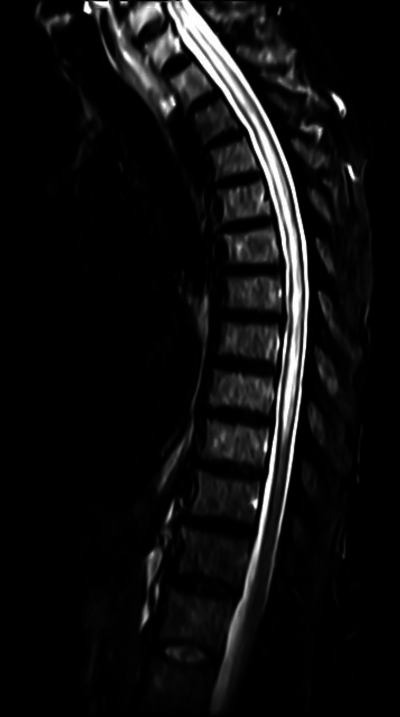
Observations: The authors report the case of a 68-year-old male suspected to have a type I dural arteriovenous fistula based on magnetic resonance angiography and angiographic evaluation. He presented with progressive myelopathy and multiple neurological symptoms exacerbated by recent trauma. A superselective angiogram of the right T10 segmental artery suggested an intradural arteriovenous fistula; however, intraoperatively, the lesion was epidural. The arterialized venous structures were obliterated, and the patient reported significant postoperative symptomatic improvement.
Lessons: This case highlights the critical importance of comprehensive imaging and cautious interpretation in the diagnosis of spinal vascular malformations. It also underscores the need for a multidisciplinary approach to ensure accurate diagnosis and effective treatment. Surgeons must be prepared for intraoperative findings that diverge from preoperative imaging to adapt surgical strategies accordingly. Furthermore, this case contributes to the evolving understanding of eAVFs, suggesting that revised imaging protocols may be required to better distinguish epidural from intradural vascular abnormalities. https://thejns.org/doi/10.3171/CASE24331.
Keywords: dural arteriovenous fistula; epidural arteriovenous fistula; neuroimaging.
Figures


References
-
- Brusko GD, Perez-Roman RJ, Tapamo H, Burks SS, Serafini AN, Wang MY. Preoperative SPECT imaging as a tool for surgical planning in patients with axial neck and back pain. Neurosurg Focus. 2019;47(6):E19. - PubMed
-
- Yamaguchi S, Takemoto K, Takeda M, et al. The position and role of four-dimensional computed tomography angiography in the diagnosis and treatment of spinal arteriovenous fistulas. World Neurosurg. 2017;103:611-619. - PubMed
-
- Takai K, Taniguchi M. Comparative analysis of spinal extradural arteriovenous fistulas with or without intradural venous drainage: a systematic literature review. Neurosurg Focus. 2012;32(5):E8. - PubMed
-
- Arnaud O, Bille F, Pouget J, Serratrice G, Salamon G. Epidural arteriovenous fistula with perimedullary venous drainage: case report. Neuroradiology. 1994;36(6):490-491. - PubMed
-
- Krings T, Mull M, Bostroem A, Otto J, Hans FJ, Thron A. Spinal epidural arteriovenous fistula with perimedullary drainage. Case report and pathomechanical considerations. J Neurosurg Spine. 2006;5(4):353-358. - PubMed
LinkOut - more resources
Full Text Sources
Research Materials
Miscellaneous
