. 2024 Oct 7;12(10):e009198.
doi: 10.1136/jitc-2024-009198.

NSGS mice humanized with cord blood mononuclear cells show sustained and functional myeloid-lymphoid representation with limited graft-versus-host disease

Affiliations

NSGS mice humanized with cord blood mononuclear cells show sustained and functional myeloid-lymphoid representation with limited graft-versus-host disease

Carla Panisello et al. J Immunother Cancer. .

Abstract

Humanized immunodeficient mice serve as critical models for investigating the functional interplay between transplanted human cells and a pre-reconstituted human immune system. These models facilitate the study of molecular and cellular pathogenic mechanisms and enable the evaluation of the efficacy and toxicity of immunotherapies, thereby accelerating their preclinical and clinical development. Current strategies rely on inefficient, long-term/delayed hematopoietic reconstitution by CD34+ hematopoietic progenitors or short-term reconstitution with peripheral blood mononuclear cells (PB-MNCs) associated with high rates of graft-versus-host disease (GvHD) and an inefficient representation of immune cell populations. Here, we hypothesized that immunologically naïve cord blood mononuclear cells (CB-MNCs) could serve as a superior alternative, providing long-lasting and functionally effective immune reconstitution. We conducted a comprehensive comparison between the non-obese diabetic (NOD).Cg-Prkdc∧ˆscid-IL2rg∧ˆtm1Wjl/SzJ (NSG) and NSG-Tg(CMV-IL3,CSF2,KITLG)∧ˆ1Eav/MloySzJ (NSGS) immunodeficient mouse models following humanization with either PB-MNCs or CB-MNCs. We assessed the engraftment dynamics of various human immune cells over time and monitored the development of GvHD in both models. For the most promising model, we extensively evaluated immune cell functionality in vitro and in vivo using sarcoma and leukemia xenografts. Humanizing NSGS mice with CB-MNCs results in a rapid, robust, and sustained representation of a diverse range of functional human lymphoid and myeloid cell populations while minimizing GvHD incidence. In this model, human immune cell populations significantly impair the growth and engraftment of sarcoma and B-cell acute lymphoblastic leukemia cells, with a significant inverse correlation between immune cell levels and tumor growth. This study establishes a fast, efficient, and reliable in vivo platform for various applications in cancer immunotherapy, particularly for exploring the complex interactions between cancer cells, immune cells, and the tumor microenvironment in vivo, prior to clinical development.

Keywords: Engraftment; Graft versus leukemia; Immunotherapy; Solid tumor; Tumor microenvironment - TME.


Conflict of interest statement

Competing interests: PM is founder of the spin-off OneChain Immunotherapeutics, which has no connection with the present research. The remaining authors declare no competing interests.

Figures
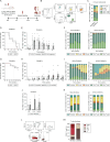
Figure 1
Figure 1. NSGS mice humanized with CB-MNCs show a rapid and sustained myeloid–lymphoid representation and low GvHD rates. (A) Schematic representation of the experimental design used to evaluate different humanization models. (B) Flow cytometry gating strategy employed to identify human immune cell subsets in mouse hematopoietic tissues. (C) PB and CB immune cell populations before MNC infusion in NSG and NSGS mice. (D) Kaplan-Meier event-free survival (EFS) curve over 10 weeks for NSG (n=9) and NSGS (n=9) mice transplanted with PB-MNCs. (E) Percentage of human immune cell (HLA-ABC+hCD45+) engraftment in the PB of NSG (n=9) and NSGS (n=9) mice transplanted with PB-MNCs monitored over a 9-week period. (F) Proportion of different human immune cell subsets within the HLA+hCD45+ human graft in the PB of NSG (n=9) and NSGS (n=9) mice transplanted with PB-MNCs. (G) Kaplan-Meier EFS over 14 weeks for NSG (n=9) and NSGS (n=12) mice transplanted with CB-MNCs. (H) Percentage of HLA-ABC+hCD45+ engraftment in the PB of NSG (n=9) and NSGS (n=9) mice transplanted with CB-MNCs monitored over a 13-week period. (I) Proportion of different human immune cell subsets within the HLA+hCD45+ human graft in the PB of NSG (n=9) and NSGS (n=12) mice transplanted with CB-MNCs. (J) Total engraftment of HLA-ABC+hCD45+ in BM, liver and spleen of NSG (n=3) and NSGS (n=3) mice transplanted with CB-MNCs. (K) Proportion of different human immune cell subsets within the HLA-ABC+hCD45+ population in BM, liver and spleen at the endpoint in NSG (n=3) and NSGS (n=3) mice transplanted with CB-MNCs. (L) Flow cytometry gating strategy used to study the T cell phenotype before PB-MNC or CB-MNC transplantation. (M) PB (n=3) and CB (n=3) T cell phenotype before transplantation. Combined results from NSGS mice humanized with PB or CB samples from three different donors are shown. Each dot represents an independent mouse. CB-MNCs, cord blood mononuclear cells; CM, central memory; EM, effector memory; EMRA, terminally differentiated effector memory cells re-expressing CD45RA; GvHD, graft-versus-host disease; N, naïve; PB-MNCs, peripheral blood mononuclear cells; SCM, stem cell memory.
Figure 2
Figure 2. Expansion of functional human immune cell populations in NSGS Mice Humanized with CB-MNCs. (A) Schematic overview of the experimental design employed to assess the ex vivo functionality of human immune cells isolated from humanized NSGS mice. Representative flow cytometry plots are shown, depicting the activation status of human immune cell populations. (B, C) Engraftment of total HLA-ABC+hCD45+ (B) and the proportion of immune populations within the HLA+hCD45+ human graft (C) before ex vivo stimulation (week 7) in PB, BM, liver, and spleen from NSGS mice transplanted with CB-MNCs. (D) Expression of CD69 and CD80 activation markers in B cells ex vivo-stimulated with CD40L and IL-4. (E) Frequency of IFNγ- and CD25-expressing CD4+T cells after ex vivo stimulation with PMA and ionomycin. (F) Frequency of IFNγ, CD25, and CD107a-expressing CD8+T cells after ex vivo stimulation with PMA and ionomycin. (G) Expression of IFNγ and CD107a in NK cells after ex vivo stimulation with PMA and ionomycin. (H) Proportion of TNFα+ and CD69+ monocytes after ex vivo stimulation with LPS. Three independent donors were used. Each dot represents data from an independent mouse. *p<0.05, **p<0.01, paired t-test. BM, bone marrow CB-MNCs, cord blood mononuclear cells; LPS, lipopolysaccharide; PB, peripheral blood; PMA, phorbol-12-miristate-13-acetate.
Figure 3
Figure 3. Human immune cell populations infiltrate subcutaneously engrafted tumors and delay tumor growth in NSGS mice humanized with CB-MNCs. (A) Schematic representation of the experimental design used to assess the tumor immunity of human immune cells against ES in vivo. (B) Kaplan-Meier overall survival (OS) curve for CB-MNCs humanized (n=20) and non-humanized (n=4) NSGS mice subcutaneously transplanted with A673 cells. (C) Weekly monitoring of tumor volume (mm3) after A673 transplantation. (D) Tumor weight (g) of independent mice at endpoint. (E) Total HLA-ABC+hCD45+ engraftment at endpoint in PB, BM, liver, spleen and subcutaneously engrafted tumors (tumor infiltration) from NSGS mice transplanted with CB-MNCs. (F) Representative immunofluorescence image of CD99+tumor cells (red) and tumor-infiltrating human CD45+immune cells (green). (G) Proportion of immune populations within the HLA+hCD45+ human graft identified at endpoint in PB, BM, liver, spleen and in subcutaneously engrafted tumors (tumor infiltration) from NSGS mice transplanted with CB-MNCs. (H–L) Correlation of tumor volume (mm3) with HLA-ABC+hCD45+ (H), Monocytes (I), B cells (J), NK cells (K) and T cells (L) in mouse PB at endpoint. Three independent donors were used. Each dot represents an independent mouse. *p<0.05, **p<0.01, ***p<0.001, ****p<0.0001, Pearson correlation test. BM, bone marrow; CB-MNCs, cord blood mononuclear cells; PB, peripheral blood.

References

    1. Allen TM, Brehm MA, Bridges S, et al. Humanized immune system mouse models: progress, challenges and opportunities. Nat Immunol. 2019;20:770–4. doi: 10.1038/s41590-019-0416-z. - DOI - PMC - PubMed
    1. Chuprin J, Buettner H, Seedhom MO, et al. Humanized mouse models for immuno-oncology research. Nat Rev Clin Oncol. 2023;20:192–206. doi: 10.1038/s41571-022-00721-2. - DOI - PMC - PubMed
    1. Capasso A, Lang J, Pitts TM, et al. Characterization of immune responses to anti-PD-1 mono and combination immunotherapy in hematopoietic humanized mice implanted with tumor xenografts. J Immunother Cancer. 2019;7:37. doi: 10.1186/s40425-019-0518-z. - DOI - PMC - PubMed
    1. Hu X, Manner K, DeJesus R, et al. Hypoimmune anti-CD19 chimeric antigen receptor T cells provide lasting tumor control in fully immunocompetent allogeneic humanized mice. Nat Commun. 2023;14:2020. doi: 10.1038/s41467-023-37785-2. - DOI - PMC - PubMed
    1. Norelli M, Camisa B, Barbiera G, et al. Monocyte-derived IL-1 and IL-6 are differentially required for cytokine-release syndrome and neurotoxicity due to CAR T cells. Nat Med. 2018;24:739–48. doi: 10.1038/s41591-018-0036-4. - DOI - PubMed

LinkOut - more resources