. 2024 Oct 8;15(1):8708.
doi: 10.1038/s41467-024-52818-0.

Coronavirus envelope protein activates TMED10-mediated unconventional secretion of inflammatory factors

Affiliations

Coronavirus envelope protein activates TMED10-mediated unconventional secretion of inflammatory factors

Lei Liu et al. Nat Commun. .

Abstract

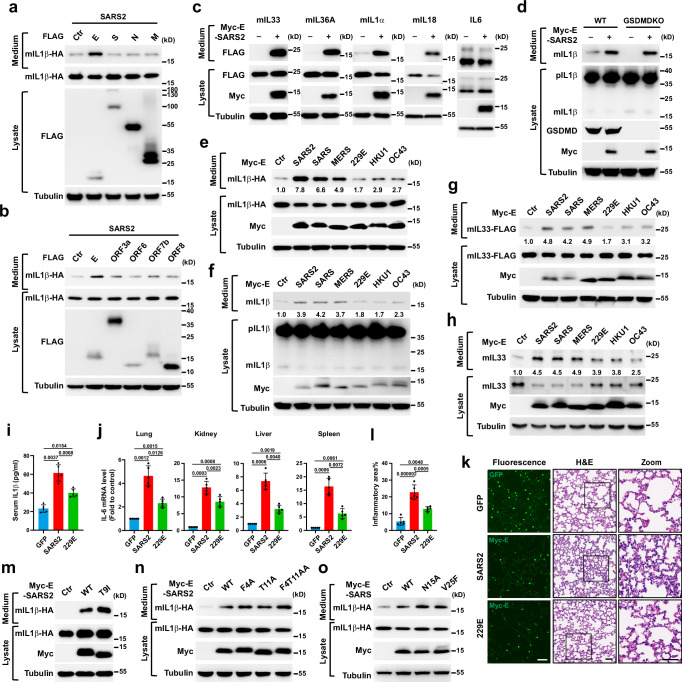
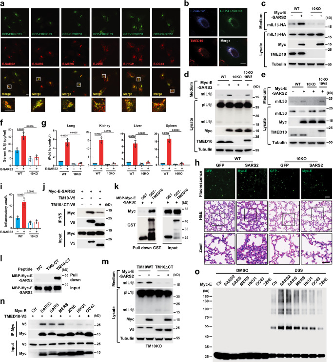
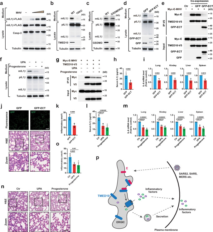
The precise cellular mechanisms underlying heightened proinflammatory cytokine production during coronavirus infection remain incompletely understood. Here we identify the envelope (E) protein in severe coronaviruses (SARS-CoV-2, SARS, or MERS) as a potent inducer of interleukin-1 release, intensifying lung inflammation through the activation of TMED10-mediated unconventional protein secretion (UcPS). In contrast, the E protein of mild coronaviruses (229E, HKU1, or OC43) demonstrates a less pronounced effect. The E protein of severe coronaviruses contains an SS/DS motif, which is not present in milder strains and facilitates interaction with TMED10. This interaction enhances TMED10-oligomerization, facilitating UcPS cargo translocation into the ER-Golgi intermediate compartment (ERGIC)-a pivotal step in interleukin-1 UcPS. Progesterone analogues were identified as compounds inhibiting E-enhanced release of proinflammatory factors and lung inflammation in a Mouse Hepatitis Virus (MHV) infection model. These findings elucidate a molecular mechanism driving coronavirus-induced hyperinflammation, proposing the E-TMED10 interaction as a potential therapeutic target to counteract the adverse effects of coronavirus-induced inflammation.


Conflict of interest statement

The authors declare no competing interests.

Figures

Fig. 1
Fig. 1. Coronavirus E protein promotes UcPS.
a, b mIL1β-HA secretion in HEK293T cells transfected with mIL1β-HA alone or together with FLAG-tagged SARS2 proteins as indicated. c Secretion of mature IL1s and IL6 in HEK293T cells with or without Myc-tagged E-SARS2 expression. d mIL1β secretion in WT or GSDMD-KO THP-1 cells with or without Myc-tagged E-SARS2 expression. e Secretion of mIL1β-HA in HEK293T cells transfected with mIL1β-HA alone or together with Myc-tagged E of indicated coronaviruses. f mIL1β secretion in THP-1 cells expressed with or without Myc-tagged E of indicated coronaviruses. g Secretion of mIL33-FLAG in HEK293T cells transfected with mIL33-FLAG alone or together with Myc-tagged E of indicated coronaviruses. h mIL33 secretion in BEAS-2B cells expressed with or without Myc-tagged E of indicated coronaviruses. il C57BL/6 WT mice injected with AAV-GFP, AAV-Myc-E-SARS2 or AAV-Myc-E-229E were challenged with 15 mg/kg LPS for 15 h and euthanized. Expression of Myc-E-SARS2, Myc-E-229E or GFP in the lung was verified by immunofluorescence (k, left panel). Serum IL1β levels were determined by ELISA in (i). IL6 mRNA levels in indicated tissues were analyzed in (j). Lung inflammation was analyzed by H&E staining (k, middle and right panel) and inflammatory area was quantified in (l). mo mIL1β-HA secretion in HEK293T cells transfected with mIL1β-HA alone or together with Myc-tagged E WT or indicated ion channel (IC) mutants of SARS2 (m, n) or SARS (o). The secretion levels of mIL1β or mIL33 were quantified based on the band intensities in the medium using Image J (eh). Data are mean ± s.d. Statistical significance was assessed using one-way ANOVA (n = 5) followed by Tukey’s multiple-comparison test. P values are indicated. Scale bars, 50 μm. Source data are provided as a Source Data file.
Fig. 2
Fig. 2. E-regulated UcPS is dependent on TMED10.
a Immunofluorescence of U2OS expressing GFP-ERGIC53 and Myc-tagged E of indicated coronaviruses. b Immunofluorescence of U2OS expressing GFP-ERGIC53, TMED10-V5 and Myc-E-SARS2. c mIL1β-HA secretion in WT and TMED10-KO HEK293T cells transfected with mIL1β-HA alone or together expressed with or without Myc-E-SARS2. d mIL1β secretion in WT and TMED10-KO THP-1 cells expressed with or without Myc-E-SARS2, TMED10-V5 or both as indicated. e mIL33 secretion in WT or TMED10-KO BEAS-2B cells in the absence or presence of Myc-E-SARS2 or TMED10-V5 expression. f–i WT and TMED10KO mice injected with AAV-GFP or AAV-Myc-E-SARS2 were challenged with 15 mg/kg LPS for 15 h and euthanized. Expression of Myc-E-SARS2 or GFP in the lung was verified by IF (h, upper panel). Serum IL1β levels were determined by ELISA in (f). IL6 mRNA levels in indicated tissues were analyzed in (g). Lung inflammation was analyzed by H&E staining (h, middle and lower panel) and inflammatory area was quantified in (i). j Co-IP using HEK293T cells with Myc-E-SARS2 and TMED10-V5 or TMED10∆CT-V5. k GST pull down analysis using GST, GST-TMED10, and MBP-Myc-E of SARS2 proteins. l Pull-down analysis using MBP-Myc-E of SARS2 protein and Ctrl, TMED6-CT or TMED10-CT peptides. m mIL1β secretion in TMED10-KO THP-1 cells transfected with or without Myc-E-SARS2 in the presence of TMED10-WT or TMED10∆CT mutant. n Co-IP using HEK293T cells with TMED10-V5 and Myc-tagged E of indicated coronaviruses. o Crosslink assay performed using HEK293T cells expressing TMED10-V5 in the absence or presence of Myc-tagged E of indicated coronaviruses. WT, Wild type; 10KO, TMED10-KO; 10V5, TMED10-V5; TM10, TMED10; TM6, TMED6; DSS, disuccinimidyl suberate. Data are mean ± s.d. Statistical significance was assessed using one-way ANOVA (n = 5) followed by Tukey’s multiple-comparison test. P values are indicated. Scale bars, 10 μm (a, b) and 50 μm (k). Source data are provided as a Source Data file.
Fig. 3
Fig. 3. E protein facilitates the membrane translocation process of TMED10-mediated UcPS.
a Diagram of in vitro translocation assay. In brief, proteoliposomes with GST-tagged Myc-E, TMED10 alone or both were incubated with recombinant mIL1β-FLAG protein at 30 °C for 1 h. Proteinase K digestion was performed to determine the amount of membrane-incorporated cargo. b In vitro membrane translocation of mIL1β-FLAG with control or GST-TMED10 proteoliposomes in the absence or presence of GST-Myc-E of SARS2. c Diagram of cell-free membrane translocation assay. In brief, membrane fraction collected from HEK293T cells expressing control (Ctrl), Myc-E-SARS2, TMED10-V5 or both after differential centrifugation was incubated with cytosol from HEK293T cells, recombinant mIL1β-FLAG plus GTP and ATPR for 1.5 h at 30 °C. After then, OptiPrep gradient ultracentrifugation was performed to remove the free proteins. Membrane floating on the top was collected and the translocated mIL1β was determined by proteinase K protection assay. d Cell-free membrane translocation of mIL1β-FLAG in the absence or presence of TMED10, Myc-E-SARS2 or both. The membrane fraction was collected from TMED10KO HEK293T cells. e Cell-free membrane translocation of mIL1β-FLAG in the absence or presence of Myc-tagged E of indicated coronaviruses. f, g Diagram of the GFP complementation assay (f). Cells expressing GFP (1–10)-TMED10 were transfected with negative control (NC) or mIL1β-GFP11 along without (Ctrl) or with Myc-tagged E of indicated coronaviruses. Complemented GFP signals were analyzed by FACS analysis and quantified in (g). Prot K, Protease K; TX-100, TritonX-100; ATPR, ATP regeneration system. Data are mean ± s.d. Statistical significance was assessed using one-way ANOVA (n = 5) followed by Tukey’s multiple-comparison test. P values are indicated. Source data are provided as a Source Data file.
Fig. 4
Fig. 4. An SS/DS motif in Es-SSC regulates TMED10-mediated UcPS.
a C-terminal partial amino acid sequence alignment of indicated coronaviruses E protein. SS/DS residues of Es-SSC are marked in red. The alternative residues of Es-MSC are marked in green. b mIL1β-HA secretion in HEK293T cells transfected with Myc-tagged E WT or AA mutant of indicated severe symptom coronaviruses. c mIL1β secretion in THP-1 cells expressed with Myc-tagged E WT or AA mutant of indicated severe symptom coronaviruses. d Co-IP using HEK293T cells with TMED10-V5 and Myc-E WT or AA mutant of indicated severe symptom coronaviruses. e Cell-free membrane translocation of mIL1β-FLAG in the absence or presence of Myc-E WT or AA mutant of SARS2. f mIL1β-HA secretion in HEK293T cells transfected with Myc-tagged E-SARS2, Myc-tagged E WT or SS mutant of indicated mild symptom coronaviruses. g mIL1β secretion in THP-1 cells expressed with Myc-tagged E-SARS2, Myc-tagged E WT or SS mutant of indicated mild symptom coronaviruses. h Co-IP using HEK293T cells with TMED10-V5 and Myc-tagged E-SARS2, Myc-tagged E WT or SS mutant of indicated mild symptom coronaviruses. i–k Cell-free membrane translocation of mIL1β-FLAG in the absence or presence of Myc-tagged E-SARS2, Myc-tagged E WT or SS mutant of indicated mild symptom coronaviruses. Prot K, Protease K; TX-100, TritonX-100. Source data are provided as a Source Data file.
Fig. 5
Fig. 5. E-TMED10 interaction is required for E-induced inflammation.
a Amino acid sequence and domains of the SARS2-E protein. The C-terminal region of SARS2-E (ECT) used to block E-TMED10 interaction is highlighted in orange. b Co-IP using HEK293T cells with TMED10-V5 and Myc-tagged E-SARS2 in the absence or presence of GFP or GFP-tagged CT of E-SARS2 (GFP-ECT). c mIL1β-HA secretion in HEK293T cells transfected with or without Myc-tagged E-SARS2, or in the absence or presence of GFP or increasing amount of GFP-ECT. d THP-1 cells were expressed with GFP or GFP-ECT followed by differentiation and mIL1β secretion determination. e Crosslink assay performed using HEK293T cells expressing TMED10-V5 in the absence or presence of Myc-E-SARS2, GFP or GFP-ECT as indicated. f Cell-free membrane translocation of mIL1β-FLAG in the absence or presence of Myc-E-SARS2 and cytosol in the presence of GFP or GFP-ECT as indicated. gj C57BL/6 mice co-injected with AAV-Myc-E-SARS2 and AAV-GFP or AAV-GFP-ECT were challenged with LPS and euthanized. Expression of Myc-E-SARS2, GFP, GFP-ECT in the lung were verified by IF (i, left panel). Serum IL1β levels were determined by ELISA (g). IL6 mRNA levels in indicated tissues were analyzed in (h). Lung inflammation was analyzed by H&E staining (i, right panel) and inflammatory area was quantified in (j). Prot K, Protease K; TX-100, TritonX-100. Data are mean ± s.d. Statistical significance was assessed using two-tailed t test (n = 5). P values are indicated. Scale bars, 50 μm. Source data are provided as a Source Data file.
Fig. 6
Fig. 6. Progesterone and UPA are inhibitors of E-induced secretion and inflammation.
a Diagram of the high-throughput compound screening system based on cargo secretion analysis by complementary NanoLuc luciferase. b Heatmap showing the average mIL1β-HiBiT secretion levels from two independent experiments by each compound normalized to control. The numbers on the left side of heatmap show the serial numbers of compounds in each row. The heatmap shows the log2 value. c Quantification of relative mIL1β secretion levels in HEK293T cells in the presence of 10 μM indicated compounds. d–e mIL1β secretion in THP-1 cells expressed with Myc-tagged E of indicated coronaviruses in the presence of 10 μM UPA (d) or Progesterone (e) for 4 h. f Co-IP using HEK293T cells with TMED10-V5 and Myc-E-SARS2 in the absence or presence of 10 μM UPA or Progesterone. g Crosslink assay performed using HEK293T cells expressing TMED10-V5 without or with Myc-E-SARS2 in the absence or presence of 10 μM UPA or Progesterone. h Cell-free membrane translocation of mIL1β-FLAG in the absence or presence of Myc-E-SARS2, UPA or Progesterone (P4) as indicated. i–k C57BL/6 mice infected with AAV-Myc-E-SARS2 were intraperitoneal injected with or without UPA (1 mg/kg) followed by challenge with LPS and euthanized. Expression of Myc-E-SARS2 in the lung was verified by immunofluorescence (k, upper panel). Serum IL1β levels were determined by ELISA in (i). IL6 mRNA levels in indicated tissues were analyzed in (j). Lung inflammation was analyzed by H&E staining (k, lower panel) and inflammatory area was quantified in (l). m Chemical structure and names of Progesterone and its analogs with (Y) or without (N) inhibitory effect on mIL1β secretion in HEK293T cells shown in (n). n Quantification of relative mIL1β secretion levels in HEK293T cells in the absence or presence of compounds in (m). DSS, disuccinimidyl suberate; Prot K, Protease K; TX-100, TritonX-100. Data are mean ± s.d. Statistical significance was assessed one-way ANOVA (n = 4) followed by Dunnett’s multiple-comparison test (c, n), two-tailed t test (n = 5) (i, j, l). P values are indicated. Scale bars, 50 μm. Source data are provided as a Source Data file.
Fig. 7
Fig. 7. E-regulates UcPS and inflammation in MHV-infected mice.
a mIL1β-FLAG secretion and Caspase-3 cleavage in HEK293T-mCC1a cells infected without or with MHV-A59 for 36 h at a MOI of 0.005, 0.05 and 0.5. b, c mIL1β secretion in MHV-A59 (MOI 0.1, 36 h) infected BMDMs from WT, TMED10 KO (b) or GSDMD KO (c) mice. d mIL1β secretion in MHV-A59 (MOI 0.1, 36 h) infected BMDMs in the presence of GFP or GFP-ECT. e Co-IP using HEK293T cells with TMED10-V5 and Myc-tagged E-MHV in the absence or presence of GFP or GFP-ECT. f mIL1β secretion in MHV-A59 (MOI 0.1, 36 h) infected BMDMs in the absence or presence of 10 μM UPA or Progesterone. g Co-IP using HEK293T cells with TMED10-V5 and Myc-E-MHV in the absence or presence of 10 μM UPA or Progesterone. h–k, IFNAR-KO mice injected with AAV-GFP or AAV-GFP-ECT were infected with MHV-A59 for 4 days and euthanized. Expression of GFP or GFP-ECT in the lung was verified by IF (j, upper panel). Serum IL1β levels were determined by ELISA (h). IL6 mRNA levels in indicated tissues was analyzed (i). Lung inflammation was analyzed by H&E staining (j, middle and lower panel) and inflammatory area was quantified (k). l–o IFNAR-KO mice infected with MHV-A59 were intraperitoneal injected with DMSO, 1 mg/kg UPA or Progesterone and euthanized. Serum IL1β levels were determined by ELISA (l). IL6 mRNA levels in indicated tissues were analyzed in (m). Lung inflammation was analyzed by H&E staining (n) and inflammatory area was quantified in (o). p A model for E-regulated UcPS. In brief, E proteins of the severe symptom coronaviruses (SARS, SARS2 and MERS) interact with TMED10 to activate the THU (TMED10-channel unconventional protein secretion)-mediated release of inflammatory cytokines. Data are mean ± s.d. Statistical significance was assessed using two-tailed t test (n = 5) (h, i, k), one-way ANOVA (n = 5) followed by Tukey’s multiple-comparison test (l, m, o). P values are indicated. Scale bars, 50μm. Source data are provided as a Source Data file.

References

    1. Tay, M. Z., Poh, C. M., Rénia, L., MacAry, P. A. & Ng, L. F. The trinity of COVID-19: immunity, inflammation and intervention. Nat. Rev. Immunol.20, 363–374 (2020). - PMC - PubMed
    1. De Wit, E., Van Doremalen, N., Falzarano, D. & Munster, V. J. SARS and MERS: recent insights into emerging coronaviruses. Nat. Rev. Microbiol.14, 523–534 (2016). - PMC - PubMed
    1. Zhang, Y.-y, Li, B.-r & Ning, B.-t The comparative immunological characteristics of SARS-CoV, MERS-CoV, and SARS-CoV-2 coronavirus infections. Front. Immunol.11, 2033 (2020). - PMC - PubMed
    1. Del Valle, D. M. et al. An inflammatory cytokine signature predicts COVID-19 severity and survival. Nat. Med.26, 1636–1643 (2020). - PMC - PubMed
    1. Xia, B. et al. SARS-CoV-2 envelope protein causes acute respiratory distress syndrome (ARDS)-like pathological damages and constitutes an antiviral target. Cell Res.31, 847–860 (2021). - PMC - PubMed

Publication types

MeSH terms

Substances

LinkOut - more resources