. 2024 Oct 8;15(1):8709.
doi: 10.1038/s41467-024-52950-x.

Structure of a Rhs effector clade domain provides mechanistic insights into type VI secretion system toxin delivery

Affiliations

Structure of a Rhs effector clade domain provides mechanistic insights into type VI secretion system toxin delivery

Brooke K Hayes et al. Nat Commun. .

Abstract

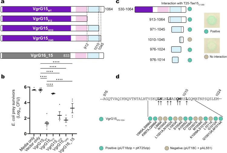
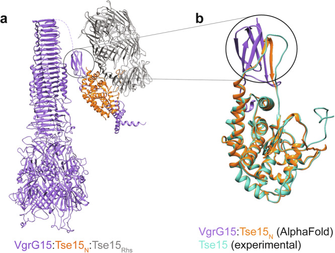
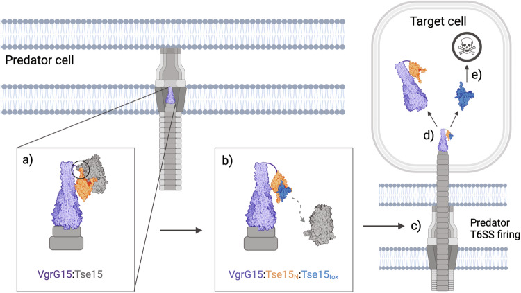
The type VI secretion system (T6SS) is a molecular machine utilised by many Gram-negative bacteria to deliver antibacterial toxins into adjacent cells. Here we present the structure of Tse15, a T6SS Rhs effector from the nosocomial pathogen Acinetobacter baumannii. Tse15 forms a triple layered β-cocoon Rhs domain with an N-terminal α-helical clade domain and an unfolded C-terminal toxin domain inside the Rhs cage. Tse15 is cleaved into three domains, through independent auto-cleavage events involving aspartyl protease activity for toxin self-cleavage and a nucleophilic glutamic acid for N-terminal clade cleavage. Proteomic analyses identified that significantly more peptides from the N-terminal clade and toxin domains were secreted than from the Rhs cage, suggesting toxin delivery often occurs without the cage. We propose the clade domain acts as an internal chaperone to mediate toxin tethering to the T6SS machinery. Conservation of the clade domain in other Gram-negative bacteria suggests this may be a common mechanism for delivery.


Conflict of interest statement

The authors declare no competing interests

Figures

Fig. 1
Fig. 1. Tse15 separates into three separate fragments that stay tightly associated during protein expression and purification.
a Construct design for production of Tse15; N-terminal clade domain is orange, Rhs domain is grey and toxin domain is blue. Purification tags and domain boundary residue numbers are indicated. b Analytical size-exclusion chromatogram showing that Tse15 elutes as a single peak. c Coomassie stained SDS-PAGE gel of purified Tse15 and domains associated with each band. Molecular weight markers (kDa) are shown on left hand side of gel. (Below) Western blots showing domain separation during purification probed with α-Strep and α-His antisera (as indicated, n = 1). Molecular weight markers (kDa) are shown on left hand side of blots. d T6SS competitive killing assays to measure the effect of replacing the Tse15 clade cleavage motif or the toxin cleavage motif with an in-frame FRT site on the ability of A. baumannii to kill vulnerable E. coli prey in a Tse15-dependent manner. Predator strains used were AB307_0294 wild-type, a ΔtssM mutant (inactive T6SS), a Δtse15 mutant, a tse15 clade cleavage mutant (tse15CC::FRT) and a tse15 toxin cleavage mutant (tse15TC::FRT). Bars represent mean of four biological replicates, error bars represent SEM. Statistical significance was determined using ANOVA with Tukey’s multiple comparisons test. ****p < 0.0001.
Fig. 2
Fig. 2. Single particle cryoEM structures of Tse15.
a Tse15 wild-type density map at a threshold of 0.75 (2.85 RMSD) where the N-terminal domain is coloured orange, Rhs cage grey and the toxin peptide(s) in blue. b Cartoon depiction of Tse15 structure coloured by domain (clade orange, Rhs grey and toxin peptides blue). Four separate unsequenced toxin peptides (106 of 195 residue CTD domain) could be modelled into the density map. To the right, Tse15 density map as mesh (ChimeraX) volume viewer at a contour of 6.45 RMSD showing fit of (c) clade-Rhs autocleavage site (S335 in grey; G333 in orange) and (e) toxin cleavage site (L1395, in grey) with toxin peptide shown in blue and Rhs domain in grey. Residues numbers are indicated. d Surface depiction of Tse15 clade and Rhs domain coloured orange and grey respectively, toxin is shown as blue cartoon. f Tse15NN density map at a threshold of 0.15 (1.2 RMSD) where the N-terminal domain is coloured orange, Rhs cage light grey and the toxin domain cyan. g Cartoon depiction of aligned Tse15 and Tse15NN where peptide toxin density is shown in blue for Tse15 and cyan for Tse15NN.
Fig. 3
Fig. 3. E. coli two-hybrid analysis of various N-terminal truncations of Tse15Δtox with VgrG15.
a Schematic representation of the regions of Tse15 fused to the C-terminal end of the T25 adenylate cyclase fragment. Each was tested for their ability to interact with one or more regions of VgrG15 fused to the C-terminal end of the T18 adenylate cyclase fragment (text at right). Region of Tse15 and derivatives shaded orange represents the clade domain, grey regions represent the Rhs domain and blue region (in Tse15 wildtype only) represents the toxin. Numbers indicate amino acid residues of wildtype Tse15 or VgrG15 each fragment represents. Interaction between a T25 fusion proteins and a T18 fusion protein is indicated with a +, no interaction is indicated by a dash. An example of a positive and negative colony can be seen in Fig. 4c. b Identified interaction sites mapped onto Tse15, with the clade domain shown in orange, the Rhs domain in grey, toxin domain in blue and the interacting regions shown in red.
Fig. 4
Fig. 4. Mapping the VgrG15 interactions with Tse15 in vivo and in vitro.
a Schematic representation of AB307-0294 wild-type VgrG15, deletion derivatives and a VgrG16:15 chimera. The purple and grey shaded area indicates conserved VgrG15 and VgrG16 domains respectively. Pink shaded region represents the VgrG15 Ig-like domain (residues 853-907) and blue shaded region indicates region predicted to form five α-helices (VgrG15 residues 933-1028). b T6SS competitive killing assays were used to determine the region of VgrG15 required for Tse15 delivery into E. coli prey cells (susceptible only to Tse15-mediated killing). The predator vgrG15 mutant was provided in trans with wild-type vgrG15, truncated vgrG15 constructs (subscript numbers indicate encoded VgrG15 amino acids), or a gene encoding a chimera VgrG16_15831-1064 protein. Bars represent mean of four biological replicates, error bars represent SEM. Statistical significance was determined using ANOVA with Tukey’s multiple comparisons test. ****p < 0.0001. c Bacterial adenylate cyclase two-hybrid analysis to measure the direct interaction of various regions of VgrG15 (fused to the C-terminal end of the T18 fragment of adenylate cyclase) with Tse15∆tox (fused to the C-terminal end of the T25 fragment of adenylate cyclase). Positive interaction between the two fusion proteins is indicated as blue/green circle while lack of interaction is indicated as cream circle (shown right of panel). d Alanine scanning of smallest region of VgrG15 still able to interact with Tse15∆tox. The ability of each alanine substituted protein (T18-VgrG976-1024 parent) to interact with Tse15∆tox was assessed using the bacterial adenylate cyclase two-hybrid system. Arrows indicate the amino acids (bold) that when substituted with alanine resulted in a failure of T18-VgrG976-1024 to interact with T25-Tse15∆tox. Representative images (minimum of three biological replicates) shown for interactions of Tse15∆tox with single and double alanine substitutions in the VgrG region of interest. Blue/green circles indicate the two recombinant proteins co-expressed are interacting. Cream coloured circles indicate no interaction was observed, showing the substituted amino acid(s) may be involved in the interaction between the two proteins.
Fig. 5
Fig. 5. Binding model for Tse15:VgrG15 prior to Rhs domain dissociation.
a Model of the full length VgrG15 homotrimer in purple interacting with the Tse15 AlphaFold2 model coloured by domain (orange clade domain and grey Rhs domain). b AlphaFold2 model of VgrG15 residues 853-907 alone (purple) interacting with the Tse15 clade (residues 20-313, orange). The cryo-EM model of Tse15 clade domain is overlaid (teal). Black circle highlights a change in N-terminal clade domain secondary structure when involved in an edge-to-edge strand interaction with VgrG15.
Fig. 6
Fig. 6. Visualisation of secretome data from A. baumannii AB307-0294.
Visualisation of secretome data from A. baumannii AB307-0294 showing the marked absence of cage peptides for Tse15 (a) and Tde16 (b). Also shown is peptide coverage for the T6SS control proteins Tae17 (a T6SS effector that does not use a Rhs cage) (c) and Hcp (a T6SS structural subunit) (d) as well as a cytoplasmic protein RpoB (e). The amino acid numbers are shown at the top of each panel. Peptide coverage and intensity are indicated for each of the two replicates of whole cell lysate (WCL) and supernatants (SUP) samples. Intensity legend is in the top right of each panel.
Fig. 7
Fig. 7. Delivery schematic for Tse15:VgrG15.
VgrG15 is depicted as purple, Tse15 is coloured by domain where clade is orange, Rhs is grey and toxin is blue. a Initial interactions between the Tse15 clade and Rhs domains and VgrG15. b Tse15 clade domain forms edge-to-edge contact interactions with VgrG15. The clade and toxin domains of Tse15 interact, triggering the Rhs dissociation. c When the Tse15 clade:toxin is bound, the T6SS fires into the target cell. d Following delivery, the toxin dissociates from protein chaperones (clade and VgrG15). The toxin can now fold independently and illicit toxic activity to cause target cell death (e). Created with BioRender.com.

References

    1. Basler, M., Pilhofer, M., Henderson, G. P., Jensen, G. J. & Mekalanos, J. J. Type VI secretion requires a dynamic contractile phage tail-like structure. Nature483, 182–186 (2012). - PMC - PubMed
    1. Leiman, P. G. et al. Type VI secretion apparatus and phage tail-associated protein complexes share a common evolutionary origin. Proc. Natl Acad. Sci. USA106, 4154–4159 (2009). - PMC - PubMed
    1. Cianfanelli, F. R., Monlezun, L. & Coulthurst, S. J. Aim, load, fire: the type VI secretion system, a bacterial nanoweapon. Trends Microbiol.24, 51–62 (2016). - PubMed
    1. Pukatzki, S., Ma, A. T., Revel, A. T., Sturtevant, D. & Mekalanos, J. J. Type VI secretion system translocates a phage tail spike-like protein into target cells where it cross-links actin. Proc. Natl Acad. Sci. USA104, 15508–15513 (2007). - PMC - PubMed
    1. Suarez, G. et al. A type VI secretion system efector protein, VgrG1, from aeromonas hydrophila that induces host cell toxicity by ADP ribosylation of actin. J. Bacteriol.192, 155–168 (2010). - PMC - PubMed

Publication types

MeSH terms

LinkOut - more resources