Clinical Trial
. 2024 Nov;131(10):1605-1612.
doi: 10.1038/s41416-024-02864-8. Epub 2024 Oct 8.

Role of 18F-FDG-PET/CT in the initial staging of very high-risk Ewing Sarcoma in a prospective multicentric Phase II Study: Is there still a place for bone marrow sampling?

Affiliations
Clinical Trial

Role of 18F-FDG-PET/CT in the initial staging of very high-risk Ewing Sarcoma in a prospective multicentric Phase II Study: Is there still a place for bone marrow sampling?

Nina Jehanno et al. Br J Cancer. 2024 Nov.

Abstract

Background: The Ewing Sarcoma Family of Tumors (ESFT) constitutes a group of rare malignancies, wherein approximately one-third of cases exhibit metastatic spread, particularly impacting prognosis when bone and/or bone marrow (BM) are involved. Primary extra-pulmonary metastatic ESFT often necessitates intensified therapeutic approaches. Accurate staging plays a pivotal role in clinical decision-making, with fluorine-18-fluorodeoxyglucose-positron emission tomography/computed tomography (PET/CT) currently serving as a non-invasive modality for assessing ESFT's BM extent.

Methods: In the French phase II COMBINAIR3 (NCT03011528) study, a comprehensive approach for patients with extra-pulmonary ESFT metastasis was evaluated. We prospectively compared the efficacy of PET/CT to BM aspiration and biopsy (BMAB) analysis in patients undergoing initial staging.

Results: Among the 42 patients analyzed (median age 14 y, 2:1 male/female ratio), 45% presented with pelvic primary tumors and 83% had bone/BM involvement at diagnosis. Our findings showed PET/CT had 100% specificity and 83.3% sensitivity in detecting initial BM involvement. Overall, PET/CT correctly classified 92.8% of patients, reaching 100% accuracy in patients identified with bone involvement, thus surpassing the standard BMAB.

Discussion: These results suggest that the conventional use of BMAB in the initial staging of high-risk ESFT patients can be omitted, promoting PET/CT as a non-invasive alternative, thus improving staging accuracy and treatment decisions in ESFT management.


Conflict of interest statement

Competing interests The authors declare no competing interests.

Figures

Fig. 1
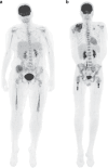
Fig. 1. Different pattern between bone metastasis and bone marrow invasion on PET/CT.
a Bone marrow involvement: intra-medullary uptake within the skeleton without evident signs of cortical destruction. Bone marrow nodular densities are discernible on the CT-scan. b Bone metastasis: Hypermetabolic focal lesions are associated with cortical destruction on CT (lytic lesion). c Distribution of infiltration patterns: bone, bone marrow (BM) and combined bone + BM. d Distribution of bone metastasis (number of sites: one lesion, 2–5, and > 5).
Fig. 2
Fig. 2. Study flowchart.
a BMAB flowchart analysis and correlation with PET/CT. b Bone marrow infiltration flowchart analysis based on PET and correlation with BMAB and MRI. Abbreviations: BM, bone marrow, BMAB, Bone Marrow Aspiration and Biopsy, PET/CT, fluorine-18-fluorodeoxyglucose (FDG)-positron emission tomography (PET)/computed tomography (CT), MRI, Magnetic resonance imaging. *Three patients were excluded due to unmet inclusion criteria (one with localized Ewing tumor after careful review of imaging and two with incorrect histological diagnosis).
Fig. 3
Fig. 3. Cases 1 and 2: Bone marrow infiltration on PET/CT.
a Case 1. Concordant positive results in BMAB with evidence of bone marrow infiltration on PET/CT. Patient with right iliac bone primary ESFT. The PET/CT revealed BM involvement characterized by a diffuse non-homogeneous infiltration in both proximal and distal skeleton regions. Both BM biopsy and cytology yielded positive results. b Case 2. Positive cytology despite negative bone marrow biopsy, with both bone and bone marrow infiltration on PET/CT. Patient with right scapula primary ESFT. The PET/CT revealed BM involvement with a diffuse non-homogeneous infiltration extending to both proximal and distal skeleton region associated with asymmetric focal uptake without evidence for cortical destruction. Although BM biopsy was negative, the cytology was positive.
Fig. 4
Fig. 4. Case 3. Discordant negative BMAB with bone marrow infiltration on PET/CT confirmed on MRI.
a Pelvic MRI corroborating bone marrow infilration, in concordance with PET/CT, in our sub-group of 7 patients with negative results BMAB. b 3D Maximum Intensity Projection (MIP) of whole-body baseline PET/CT c Fused PET/CT and d CT scans assessing nodular intra-medullar infiltration in both femurs. e Coronal T1 gadolinium-enhanced MRI of the pelvis and f with fat saturation, is consistent with PET/CT findings of BM infiltration.
Fig. 5
Fig. 5. Illustration of changes observed in femoral BM from birth to adulthood and its representation in our cohort.
a Bone marrow composition changes across the lifespan (personal illustration). The composition of BM undergoes dynamic changes throughout an individual’s life, marked by the conversion of hematopoietic red marrow into fatty yellow marrow. Initially, at birth, the medullary cavity exclusively contains hematopoietic red marrow. The conversion process initiates in the diaphysis, with a bidirectional progression towards the metaphysis, greater distally. In young adults, predominant red marrow presence is observed in the proximal metaphysis of long bones. b Bone marrow infiltration patterns on baseline PET/CT across age groups in our population. Younger children exhibit a higher prevalence of involvement of distal metaphysis, while older young adults predominantly display infiltration in the proximal femoral metaphysis, consistent with hematopoietic red marrow maturation.

References

    1. Fletcher CDM, World Health Organization, International Agency for Research on Cancer, editors. WHO classification of tumours of soft tissue and bone. 4th ed. Lyon: IARC Press; 2013. 468 (World Health Organization classification of tumours).
    1. Dockhorn-Dworniczak B, Schäfer KL, Dantcheva R, Blasius S, Winkelmann W, Strehl S, et al. Diagnostic value of the molecular genetic detection of the t(11;22) translocation in Ewing’s tumours. Virchows Arch Int J Pathol. 1994;425:107–12. - PubMed
    1. Delattre O, Zucman J, Melot T, Garau XS, Zucker JM, Lenoir GM, et al. The Ewing family of tumors–a subgroup of small-round-cell tumors defined by specific chimeric transcripts. N. Engl J Med. 1994;331(Aug 4):294–9. - PubMed
    1. Kovar H, Dworzak M, Strehl S, Schnell E, Ambros IM, Ambros PF, et al. Overexpression of the pseudoautosomal gene MIC2 in Ewing’s sarcoma and peripheral primitive neuroectodermal tumor. Oncogene. 1990;5(Jul):1067–70. - PubMed
    1. Delattre O, Zucman J, Plougastel B, Desmaze C, Melot T, Peter M, et al. Gene fusion with an ETS DNA-binding domain caused by chromosome translocation in human tumours. Nature. 1992;359:162–5. - PubMed

Publication types

Substances

LinkOut - more resources