. 2024 Oct 8;7(1):1279.
doi: 10.1038/s42003-024-06999-5.

USP24 promotes autophagy-dependent ferroptosis in hepatocellular carcinoma by reducing the K48-linked ubiquitination of Beclin1

Affiliations

USP24 promotes autophagy-dependent ferroptosis in hepatocellular carcinoma by reducing the K48-linked ubiquitination of Beclin1

Jiahui Cao et al. Commun Biol. .

Abstract

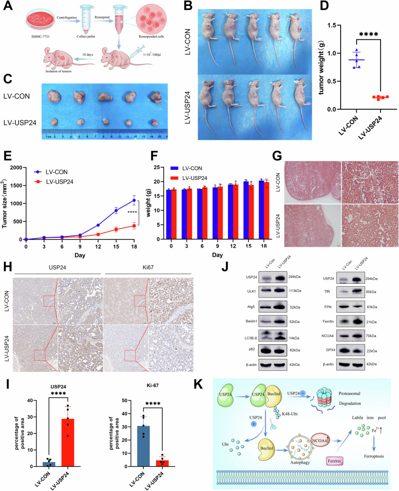
Ubiquitination is a post-translational modification (PTM), which is critical to maintain cell homeostasis. Ubiquitin-specific protease 24 (USP24) plays roles in various diseases, the mechanisms by which USP24 regulates hepatocellular carcinoma (HCC) remain poorly understood. In this study, USP24 is found to be significantly downregulated in HCC. Knocking down USP24 promotes HCC proliferation and migration, whereas USP24 overexpression inhibits HCC in vitro and in vivo. The endogenous interaction between USP24 and Beclin1 is confirmed. Mechanically, USP24 delays Beclin1 degradation by reducing its K48-linked ubiquitination, the effects of overexpressing USP24 on HCC proliferation can be partially reversed by silencing Beclin1. We find that increased autophagy is accompanied by ferroptosis in USP24 overexpressed HCC cells and USP24 increases the susceptibility of HCC to sorafenib. Collectively, this study highlights the critical role of USP24 in regulating autophagy-dependent ferroptosis by decreasing Beclin1 ubiquitination, suggesting that targeting USP24 may be a strategy for treating HCC.


Conflict of interest statement

The authors declare no competing interests.

Figures

Fig. 1
Fig. 1. Low expression of USP24 protein is correlated with poor prognosis in HCC patients.
A The mRNA level of USP24 in normal liver cell and HCC cell lines was measured by qRT-PCR (n = 3). B The expression level of USP24 protein in normal liver cell and HCC cell lines was measured by western blot. C Immunohistochemistry assay was performed in paracancerous tissue and paired carcinoma tissue microarray (n = 70). Representative images of USP24 in normal tissues, HCC tissues with different levels of USP24 are shown. Scale bar, 20 μm. D Quantification of the USP24 expression in paracancerous tissues and carcinoma tissues. E Kaplan-Meier curves from patients with HCC expressing low (n = 39) and high (n = 31) USP24 from the tissue microarray. *P < 0.05, **P < 0.01, ***P < 0.001.
Fig. 2
Fig. 2. Inhibition of USP24 promotes proliferation and migration of HCC cells.
A and B The knockdown efficiency of USP24 by siRNA was measured by qRT-PCR and western blot (n = 3). C CCK-8 assays were conducted to determine the viability of SMMC-7721 and HCCLM3 cells after knockdown of USP24 (n = 3). D–E Colony formation assays of HCC cells treated with si-USP24 or si-NC were performed to evaluate the ability of proliferation. The clone number was counted using ImageJ (n = 3). F Migration abilities of SMMC-7721 and HCCLM3 cells after knockdown of USP24 were evaluated with wound healing assay. Scale bar, 1 mm. G–H Transwell assays were performed to detect the ability of migration. The migration rate was calculated (n = 3). Scale bar, 100 μm. *P < 0.05, **P < 0.01, ***P < 0.001, ****P < 0.0001.
Fig. 3
Fig. 3. USP24 increased the level of autophagy.
A The relationship of USP24 expression with autophagy markers was investigated via Gene Expression Profiling Interactive Analysis (GEPIA). B qRT-PCR was performed to measure the mRNA expression of ULK1, Atg5 and Beclin1 after silencing USP24 (n = 3). C qRT-PCR was performed to measure the mRNA expression of ULK1, Atg5 and Beclin1 after overexpressing USP24 (n = 3). D The expression of ULK1, Atg5, Beclin1, LC3B-II and p62 proteins in USP24 knockdown HCC cells were measured by western blot. E The expression of autophagy related proteins after overexpressing USP24 was detected by western blot. F Transmission electron microscopy (TEM) was used to detect the formation of autophagic structures. Red arrows denote representative autolysosome. Scale bar, 5 μm (left), 500 nm (right). G The number of autophagic vesicles was counted and presented in the image (n = 3). *P < 0.05, **P < 0.01, ***P < 0.001.
Fig. 4
Fig. 4. USP24 interacts with Beclin1 and increases the stability of Beclin1 by decreasing its K48-linked ubiquitination.
A, B Reciprocal Co-IP experiments were performed to detect the interaction between USP24 and Beclin1. An IgG antibody was used as the control. C The cell double immunofluorescence assay was carried out using anti-USP24 and anti-Beclin1 antibodies. The colocalization of USP24 (red) and Beclin1 (green) was observed under a confocal microscope (n = 3). Scale bar, 10 μm. D The colocalization was evaluated by Mander’s colocalization coefficients in ten regions of interest (n = 10). E SMMC-7721 cells were treated with 10 μg/ml MG132 before harvesting. An anti-Beclin1 antibody was used to immunoprecipitate the Beclin1 protein, then an anti-Ubi antibody was used for western blot. F, G The levels of K48 and K63-linked ubiquitination were measured by western blot. H–K Control and USP24 overexpressed HCCLM3 and SMMC-7721 cells were treated with CHX (100 μg/ml) for indicated time. The expression level of Beclin1 was determined by western blot (n = 3). *P < 0.05, **P < 0.01, ***P < 0.001, ****P < 0.0001.
Fig. 5
Fig. 5. USP24 inhibits the proliferation and migration of HCC cells.
A, B The overexpression efficiency of USP24 by lentivirus was measured by western blot and qRT-PCR (n = 3). C CCK-8 assays were conducted to determine the viability of SMMC-7721 and HCCLM3 cells after overexpressing USP24 (n = 3). D–E Colony formation assays of HCC cells treated with LV-CON or LV-USP24 were performed to evaluate the ability of proliferation. The clone number was counted (n = 3). F Migration abilities of SMMC-7721 and HCCLM3 cells after overexpression of USP24 were evaluated by wound healing assay. Scale bar, 1 mm. G-H Transwell assays were performed to detect the ability of migration, and the migration rate was calculated (n = 3). Scale bar, 100 μm. **P < 0.01, ***P < 0.001, ****P < 0.0001.
Fig. 6
Fig. 6. Knockdown of Beclin1 rescues the cell proliferation and migration viability caused by overexpressing USP24.
AC The cells overexpressed USP24 were treated with Bafilomycin A1 (10 nM) for 24 h. A CCK8 assay was used to detect the cell viability (n = 5). B–C Transwell assay was performed to evaluate the migration ability of HCC cells, and the migration rate was calculated (n = 3). D The efficiency of Beclin1 knockdown was measured by western blotting. E CCK8 assay was performed to determine the cell viability after USP24 overexpression or Beclin-1 knockdown (n = 3). F–G Transwell assays of HCC cells with USP24 overexpression or Beclin1 knockdown were performed to evaluate the ability of migration, and the migration rate was calculated (n = 3). *P < 0.05, **P < 0.01, ***P < 0.001, ****P < 0.0001.
Fig. 7
Fig. 7. USP24 promotes autophagy-dependent ferroptosis in HCC cells.
A USP24 overexpressed SMMC-7721 and HCCLM3 cells were treated with Fer-1 (2 μM), Nec-1 (1 μM) and z-VAD-fmk (10 μM) separately. The cell viability was determined by CCK8 assay (n = 3). B Erastin (20 μM) was used to induce ferroptosis for 24 h, simultaneously treated with Fer-1 (2 μM), Nec-1 (1 μM) and z-VAD-fmk (10 μM). Cell viability was determined by CCK8 assay (n = 3). C Intracellular Fe2+ was detected with the FerroOrange probe (1 μM) under an inverted fluorescence microscope. Scale bar, 100 μm. D The Fe2+ fluorescence intensity was quantified by ImageJ (n = 3). E The intracellular Fe2+ was measured by colorimetric method (n = 3). F–G The mRNA and protein expression of NCOA4, TfR, FPN, ferritin and GPX4 after overexpressing USP24 was measured by qRT-PCR and WB (n = 3). H The level of GSH in HCC cells transfected with LV-CON or LV-USP24 vectors (n = 3). I The level of MDA in HCC cells transfected with LV-CON or LV-USP24 vectors (n = 3). J Control and USP24 overexpressed SMMC-7721 cells were treated with Sorafenib (0, 5, 10, 15, 20 and 25 μM) for 48 h, the cell viabilities were determined by CCK8 assay (n = 3). *P < 0.05, **P < 0.01, ***P < 0.001, ****P < 0.0001.
Fig. 8
Fig. 8. USP24 inhibits HCC growth in vivo.
A The process of constructing subcutaneous tumor models in nude mice. B–C Image of subcutaneous tumors in control and USP24 overexpressed groups (n = 5). D The tumor weight is presented as the mean ± standard deviation (n = 5). E Tumor volumes were calculated every 3 days after injection. F The body weight of nude mice during this experiment (n = 5). G Representative images of H&E stained tumor tissues. H Representative images of IHC stained USP24 and Ki-67 in control tissues or USP24 overexpressed xenograft tumor tissues. I The quantification of USP24 and Ki-67 positive cells (n = 5). J Western blot was performed to detect the expression of proteins associated with autophagy and ferroptosis. K Graphical summary of USP24 regulated autophagy-dependent ferroptosis in HCC. ****P < 0.0001.

References

    1. Sung, H. et al. Global Cancer Statistics 2020: GLOBOCAN Estimates of Incidence and Mortality Worldwide for 36 Cancers in 185 Countries. CA Cancer J. Clin.71, 209–249 (2021). - PubMed
    1. McGlynn, K. A., Petrick, J. L. & El-Serag, H. B. Epidemiology of Hepatocellular Carcinoma. Hepatol. (Baltim., Md.)73, 4–13 (2021). - PMC - PubMed
    1. Forner, A., Reig, M. & Bruix, J. Hepatocellular carcinoma. Lancet391, 1301–1314 (2018). - PubMed
    1. Llovet, J. M. et al. Immunotherapies for hepatocellular carcinoma. Nat. Rev. Clin. Onco l19, 151–172 (2022). - PubMed
    1. Huang, A., Yang, X. R., Chung, W. Y., Dennison, A. R. & Zhou, J. Targeted therapy for hepatocellular carcinoma. Signal Transduct. Target Ther.5, 146 (2020). - PMC - PubMed

MeSH terms