. 2024 Nov;43(48):3489-3497.
doi: 10.1038/s41388-024-03186-y. Epub 2024 Oct 8.

Inhibition and degradation of NRAS with a pan-NRAS monobody

Affiliations

Inhibition and degradation of NRAS with a pan-NRAS monobody

Michael Whaby et al. Oncogene. 2024 Nov.

Abstract

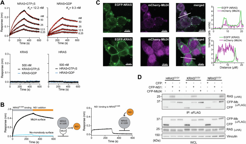
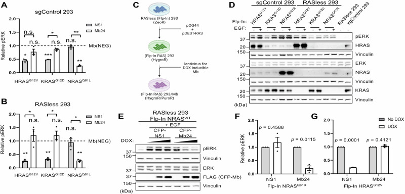
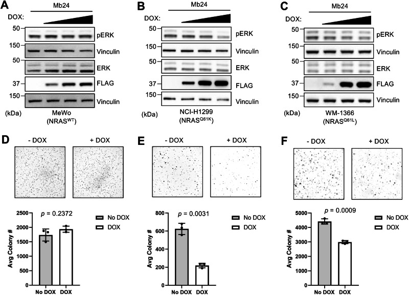
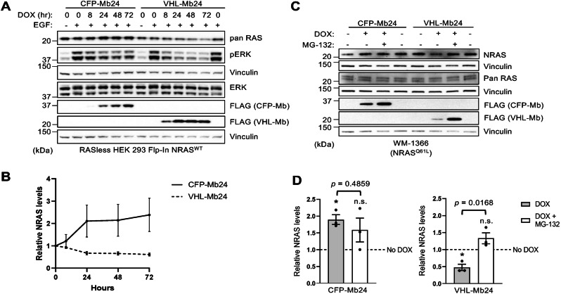
The RAS family GTPases are the most frequently mutated oncogene family in human cancers. Activating mutations in either of the three RAS isoforms (HRAS, KRAS, or NRAS) are found in nearly 20% of all human tumors with NRAS mutated in ~25% of melanomas. Despite remarkable advancements in therapies targeted against mutant KRAS, NRAS-specific pharmacologics are lacking. Thus, development of inhibitors of NRAS would address a critical unmet need to treating primary tumors harboring NRAS mutations as well as BRAF-mutant melanomas, which frequently develop resistance to clinically approved BRAF inhibitors through NRAS mutation. Building upon our previous studies with the monobody NS1 that recognizes HRAS and KRAS but not NRAS, here we report the development of a monobody that specifically binds to both GDP and GTP-bound states of NRAS and inhibits NRAS-mediated signaling in a mutation-agnostic manner. Further, this monobody can be formatted into a genetically encoded NRAS-specific degrader. Our study highlights the feasibility of developing NRAS selective inhibitors for therapeutic efforts.


Conflict of interest statement

Competing interests: JPO, AK and SK are listed as inventors on a patent application on Monobodies targeting the nucleotide-free state of RAS files by the Medical University of South Carolina and New York University (No. 62/862,924). KWT, AK, and SK are listed as inventors on a patent application on mutant RAS targeting Monobodies filed by New York University (Application No. 63/121,903). AK and SK are listed as inventors on issued and pending patents on Monobody technology filed by The University of Chicago (US Patent 9512199 B2 and related pending applications), and on a pending patent on NRAS-selective monobodies (WO2023192915A1). SK is a co-founder and holds equity in Aethon Therapeutics; is a co-founder and holds equity in Revalia Bio; was an SAB member and received consulting fees from Black Diamond Therapeutics; has received research funding from Aethon Therapeutics, Argenx BVBA, Black Diamond Therapeutics, and Puretech Health. The other authors declare no competing interests. The other authors declare no competing interests. Ethics approval and consent to participate: This study did not use any human subjects, vertebrate animals or identifiable images from human research patients.

Figures

Fig. 1
Fig. 1. Mb24 specifically interacts with NRAS.
A Biolayer interferometry (BLI) sensorgrams of RAS isotypes loaded with either GTPγS or GDP to Mb24 immobilized on a sensor tip. The KD values shown are from global fitting of a 1:1 binding model. N.D. not determined due to weak binding. B Mb24 and NS1 share an overlapping epitope. BLI sensorgram of immobilized Mb24 binding to NRAS(K135R)•GDP binding followed by the addition of NS1 (left graph). NS1 did not bind to the NRAS(K135R) precomplexed with Mb24. NS1 does bind to immobilized NRAS(K135R) in the absence of Mb24 (right graph). C Colocalization of mCherry-tagged Mb24 with EGFP-tagged KRAS4B or NRAS in cotransfected HEK 293 cells. The graphs show the fluorescence intensity profiles across the microscopy images. D Coimmunoprecipitation of Mbs from HEK 293 cells cotransfected with FLAG-tagged CFP alone, CPF-NS1, or CFP-Mb24 and HA-tagged HRASG12V, KRASG12V, or NRASG12V. Top panel, αFLAG immunoprecipitates were probed with αFLAG and αHA antibodies. Bottom panels, whole cell lysates (WCL) were probed with the indicated antibodies.
Fig. 2
Fig. 2. Effect of Mb24 on NRAS signaling in cells.
A Quantification of pERK/ERK in parental HEK 293 cells as well as (B) RASless HEK 293 cells cotransfected with CFP-FLAG-tagged Mb(NEG), NS1 or Mb24 and HRASG12V, KRASG12D, or NRASQ61L. All results were normalized to Mb(NEG). All experiments were repeated at least three times (n = 3) and results quantified using Welch’s t-test; error bars represent SEM. (***p < 0.0005, **p < 0.005, and *p < 0.05, n.s. not significant). Asterisks under brackets are pERK/ERK values from NS1 or Mb24 compared to Mb(NEG) while those above brackets are comparisons of NS1 versus Mb24. C Illustration of the workflow to generate RASless HEK 293 cells which stably express different RAS isoforms/mutants and/or different Mbs. D Western blot of control HEK 293 cells (sgControl 293) and RASless HEK 293 cells, which stably express different RAS isoforms/mutants, stimulated with ± EGF (20 ng/mL for 5 min). E Effect on pERK of DOX-induced (0, 1, and 10 μg/mL DOX) CFP-FLAG-tagged NS1 or Mb24 in EGF-stimulated RASless HEK 293 (Flp-In wild-type NRAS) cells. F Quantification of Mb effect on pERK/ERK in RASless HEK 293 (Flp-In NRASQ61R) and (G) RASless HEK 293 (Flp-In HRASG12V) (n = 3). Results quantified using Welch’s t-test; error bars represent SEM.
Fig. 3
Fig. 3. Mb24 activity in tumor cell lines.
DOX titration (0, 0.1, 1.0, 10.0 μg/mL) of stable Mb24-expressing tumor cell lines [A MeWo (wild-type NRAS), B NCI-H1299 (NRASQ61K), C WM-1366 (NRASQ61L)]. ERK phosphorylation was quantified using densitometry measurements of pERK/ERK from Western blots (Supplementary Figs. 9 and 10) (n = 3). DF Soft agar assays to analyze the effect of DOX-induced Mb24 expression on anchorage-independent growth of indicated tumor cell lines (n = 3). Colonies from −DOX and +DOX samples were quantified using ImageJ and Welch’s t-test was used to compare −DOX and +DOX samples; error bars represent SD.
Fig. 4
Fig. 4. Mechanism of action of NRAS inhibition by Mb24 in live cells.
A Illustration of the NanoBiT assay used to assess PPIs in live cells. B Effect of NS1 (gray) and Mb24 (white) on the interaction of SmBiT-CRAF with LgBiT-KRASG12V or LgBiT-NRASQ61R. C Effect of NS1 and Mb24 on SmBiT-CRAF and LgBiT-BRAF interaction in cells cotransfected with HA-tagged KRASG12V or NRASQ61R. D Effect of NS1 or Mb24 on LgBiT- and SmBiT-tagged KRASG12V or NRASQ61R to assess the effect of these Mbs on RAS self-association. All experiments were repeated four times (n = 4), normalized to a control Mb (Mb(NEG), represented by the dotted line), and analyzed using a paired t-test to compare the effects of NS1 versus Mb24; error bars represent SD.
Fig. 5
Fig. 5. Inducible degradation of NRAS by VHL-Mb24.
A Expression of CFP-Mb24 or VHL-Mb24 were chemically induced in RASless HEK 293 (Flp-In NRASWT) cells for the indicated time points and then cells were treated with EGF (20 ng/ml) for 5 min before lysate preparation. B Quantification of NRAS levels from (A). NRAS levels were normalized to vinculin loading control, and all time points were normalized to 0 h of DOX treatment. NRAS levels from each time point were compared to 0-h time points to determine statistical significance using Welch’s t-test. Only the 48- and 72-h timepoints for VHL-Mb24 with DOX were statistically different (p = 0.046 and 0.025, respectively). Experiments were repeated three times (n = 3); error bars represent SEM. C Proteasome inhibition with MG-132 rescues NRAS protein levels in WM-1366 cells which express CFP-Mb24 or VHL-Mb24 upon DOX induction. D NRAS levels from (C) were normalized to vinculin and all values for either CFP-Mb24 or VHL-Mb24 were normalized to No DOX (dotted line). Results quantified were using Welch’s t-test; error bars represent SEM. (*p < 0.05, n.s. not significant). Asterisks under brackets represent a significant difference for normalized NRAS values from DOX or DOX + MG-132 compared to No DOX while values above brackets represent comparison of DOX versus DOX + MG-132. Experiments were repeated three times (n = 3).

References

    1. Karnoub AE, Weinberg RA. Ras oncogenes: split personalities. Nat Rev Mol Cell Biol. 2008;9:517–31. - PMC - PubMed
    1. Prior IA, Lewis PD, Mattos C. A comprehensive survey of Ras mutations in cancer. Cancer Res. 2012;72:2457–67. - PMC - PubMed
    1. Prior IA, Hood FE, Hartley JL. The frequency of ras mutations in cancer. Cancer Res. 2020;80:2969–74. - PMC - PubMed
    1. Heppt MV, Siepmann T, Engel J, Schubert-Fritschle G, Eckel R, Mirlach L, et al. Prognostic significance of BRAF and NRAS mutations in melanoma: a German study from routine care. BMC Cancer. 2017;17:536. - PMC - PubMed
    1. Heidorn SJ, Milagre C, Whittaker S, Nourry A, Niculescu-Duvas I, Dhomen N, et al. Kinase-dead BRAF and oncogenic RAS cooperate to drive tumor progression through CRAF. Cell. 2010;140:209–21. - PMC - PubMed