

Novel deep learning radiomics nomogram-based multiparametric MRI for predicting the lymph node metastasis in rectal cancer: A dual-center study
- PMID: 39379733
- PMCID: PMC11461781
- DOI: 10.1007/s00432-024-05986-x
Novel deep learning radiomics nomogram-based multiparametric MRI for predicting the lymph node metastasis in rectal cancer: A dual-center study
Abstract
Purpose: To develop and evaluate a nomogram that integrates clinical parameters with deep learning radiomics (DLR) extracted from Magnetic Resonance Imaging (MRI) data to enhance the predictive accuracy for preoperative lymph node (LN) metastasis in rectal cancer.
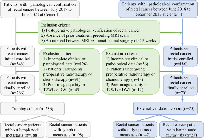
Methods: A retrospective analysis was conducted on 356 patients diagnosed with rectal cancer. Of these, 286 patients were allocated to the training set, and 70 patients comprised the external validation cohort. Preprocessed T2-weighted and diffusion-weighted imaging performed preoperatively facilitated the extraction of DLR features. Five machine learning algorithms-k-nearest neighbor, light gradient boosting machine, logistic regression, random forest, and support vector machine-were utilized to develop DLR models. The most effective algorithm was identified and used to establish a clinical DLR (CDLR) nomogram specifically designed to predict LN metastasis in rectal cancer. The performance of the nomogram was evaluated using receiver operating characteristic curve analysis.
Results: The logistic regression classifier demonstrated significant predictive accuracy using the DLR signature, achieving an Area Under the Curve (AUC) of 0.919 in the training cohort and 0.778 in the external validation cohort. The integrated CDLR nomogram exhibited robust predictive performance across both datasets, with AUC values of 0.921 in the training cohort and 0.818 in the external validation cohort. Notably, it outperformed both the clinical model, which had AUC values of 0.770 and 0.723 in the training and external validation cohorts, respectively, and the stand-alone DLR model.
Conclusion: The nomogram derived from multiparametric MRI data, referred to as the CDLR model, demonstrates strong predictive efficacy in forecasting LN metastasis in rectal cancer.
Keywords: Deep learning; Lymph node metastasis; Nomogram; Radiomics; Rectal cancer.
© 2024. The Author(s).
Conflict of interest statement
The authors declare that they have no conflict of interest.
The authors declare no competing interests.
Figures


References
-
- Beets-Tan RGH, Lambregts DMJ, Maas M et al (2017) Magnetic resonance imaging for clinical management of rectal cancer: updated recommendations from the 2016 European Society of Gastrointestinal and Abdominal Radiology (ESGAR) consensus meeting. Eur Radiol 28(4):1465–1475. 10.1007/s00330-017-5026-2 - DOI - PMC - PubMed
Publication types
MeSH terms
Grants and funding
LinkOut - more resources
Full Text Sources
