. 2024 Oct 8;81(1):428.
doi: 10.1007/s00018-024-05461-3.

Functional diversification process of opsin genes for teleost visual and pineal photoreceptions

Affiliations

Functional diversification process of opsin genes for teleost visual and pineal photoreceptions

Chihiro Fujiyabu et al. Cell Mol Life Sci. .

Abstract

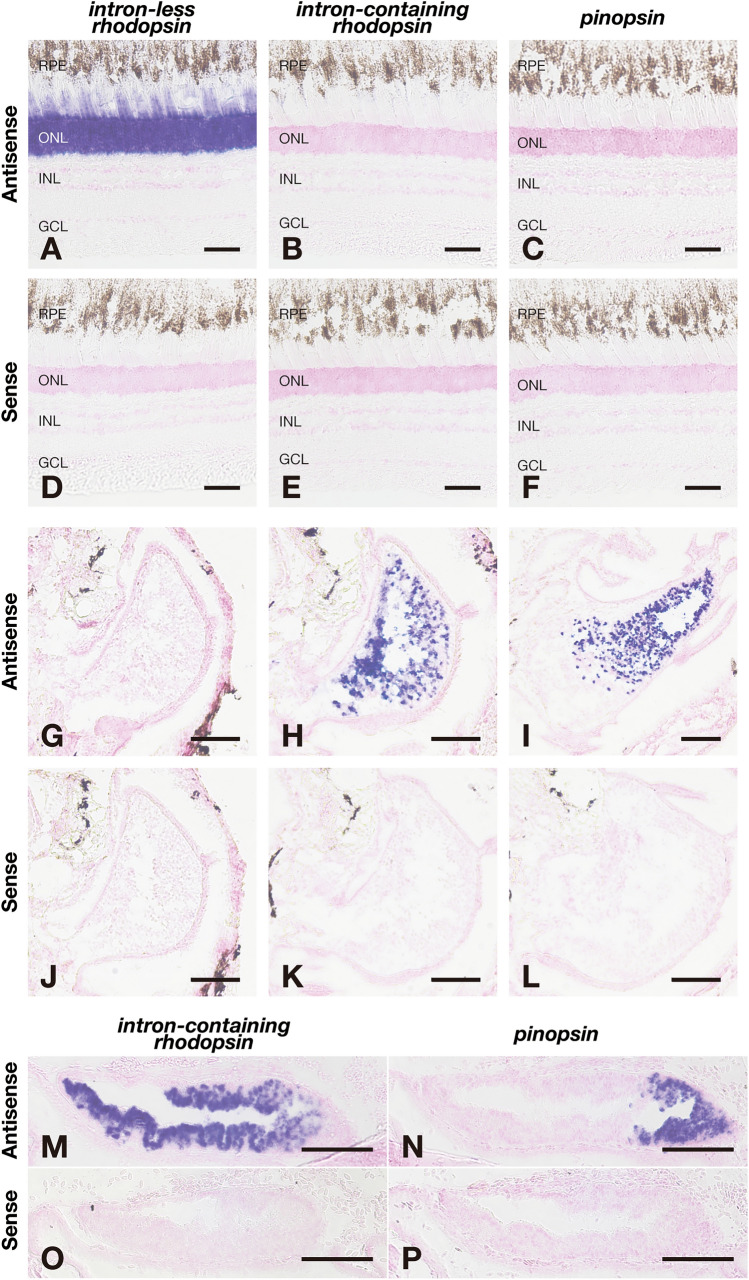
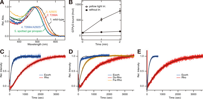
Most vertebrates have a rhodopsin gene with a five-exon structure for visual photoreception. By contrast, teleost fishes have an intron-less rhodopsin gene for visual photoreception and an intron-containing rhodopsin (exo-rhodopsin) gene for pineal photoreception. Here, our analysis of non-teleost and teleost fishes in various lineages of the Actinopterygii reveals that retroduplication after branching of the Polypteriformes produced the intron-less rhodopsin gene for visual photoreception, which converted the parental intron-containing rhodopsin gene into a pineal opsin in the common ancestor of the Teleostei. Additional analysis of a pineal opsin, pinopsin, shows that the pinopsin gene functions as a green-sensitive opsin together with the intron-containing rhodopsin gene for pineal photoreception in tarpon as an evolutionary intermediate state but is missing in other teleost fishes, probably because of the redundancy with the intron-containing rhodopsin gene. We propose an evolutionary scenario where unique retroduplication caused a "domino effect" on the functional diversification of teleost visual and pineal opsin genes.

Keywords: Opsin; Pineal gland; Pinopsin; Retina; Retroduplication; Rhodopsin.


Conflict of interest statement

The authors declare no competing interests.

Figures

Fig. 1
Fig. 1
The synteny analysis of rhodopsin and pinopsin genes in the Actinopterygii. The synteny block of orthologous genes flanking the intron-containing rhodopsin gene (blue triangle with vertical lines), the intron-less rhodopsin gene (pink triangle), and the pinopsin gene (light green triangle) in actinopterygian species. The genes flanking the opsin loci are shown by white triangles. Gene names are indicated above the coelacanth (Latimeria chalumnae) genes, intraflagellar transport 122 (ift122) (gene number 1 shown by white triangle), H1.8 linker histone (h1-8) (gene number 2), plexin D1 (plxnd1) (gene number 3), prickle planar cell polarity protein 2 (prickle2) (gene number 4), ADAM metallopeptidase with thrombospondin type 1 motif 9 (adamts9) (gene number 5) and membrane-associated guanylate kinase, WW and PDZ domain containing 1 (magi1) (gene number 6), double C2 domain beta (doc2b) (gene number 7), L-asparaginase (L-ASNase) (gene number 8), and cytosolic arginine sensor for mTORC1 subunit 2 (castor2) (gene number 9). In Atlantic tarpon (M. atlanticus), regarding the two intron-less rhodopsin genes, the gene flanking adamts9 is found in the conserved synteny block, whereas the genome region including the other gene is deleted. Detailed gene information is shown in Supplementary file 2
Fig. 2
Fig. 2
Distribution of mRNA of the intron-less and intron-containing rhodopsin genes in the retina and the pineal gland of Australian bonytongue. A-D, Distribution of the transcripts of Australian bonytongue intron-less rhodopsin gene (A, C) and intron-containing rhodopsin gene (B, D) in the retina. These sections were hybridized with antisense probes (A, B) or corresponding sense probes (C, D). Scale bar: 50 μm. Abbreviations: RPE, retinal pigment epithelium; ONL, outer nuclear layer; INL, inner nuclear layer; GCL, ganglion cell layer. EH, Distribution of the transcripts of Australian bonytongue intron-less rhodopsin gene (E, G) and intron-containing rhodopsin gene (F, H) in the transverse sections of the pineal gland. These sections were hybridized with antisense probes (E, F) or corresponding sense probes (G, H). Scale bar: 100 μm
Fig. 3
Fig. 3
Distribution of mRNA of the intron-less and intron-containing rhodopsin genes in the retina and the pineal gland of Japanese eel. AF, Distribution of the transcripts of Japanese eel intron-less rhodopsin genes, namely freshwater type rhodopsin gene (fw-rho) (A, D) and deep-sea type rhodopsin gene (ds-rho) (B, E), and intron-containing rhodopsin gene (C, F) in the retina. These sections were hybridized with antisense probes (AC) or corresponding sense probes (DF). Scale bar: 50 μm. Abbreviations: RPE, retinal pigment epithelium; ONL, outer nuclear layer; INL, inner nuclear layer; GCL, ganglion cell layer. GL, Distribution of the transcripts of Japanese eel fw-rho gene (G, J), ds-rho gene (H, K), and intron-containing rhodopsin gene (I, L) in the transverse sections of the pineal gland. These sections were hybridized with antisense probes (GI) or corresponding sense probes (JL). Scale bar: 50 μm
Fig. 4
Fig. 4
Distribution of mRNA of the intron-less and intron-containing rhodopsin genes and pinopsin gene in the retina and the pineal gland of Atlantic tarpon. AF, Distribution of the transcripts of Atlantic tarpon intron-less rhodopsin gene (A, D), intron-containing rhodopsin gene (B, E), and pinopsin gene (C, F) in the retina. These sections were hybridized with antisense probes (AC) or corresponding sense probes (DF). Scale bar: 50 μm. Abbreviations: RPE, retinal pigment epithelium; ONL, outer nuclear layer; INL, inner nuclear layer; GCL, ganglion cell layer. GL, Distribution of the transcripts of Atlantic tarpon intron-less rhodopsin gene (G, J), intron-containing rhodopsin gene (H, K), and pinopsin gene (I, L) in the transverse sections of the pineal gland. These sections were hybridized with antisense probes (GI) or corresponding sense probes (JL). Scale bar: 100 μm. MP, Distribution of the transcripts of Atlantic tarpon intron-containing rhodopsin gene (M, O) and pinopsin gene (N, P) in the sagittal sections of the pineal gland. Rostral is to the left, and dorsal is up. These sections were hybridized with antisense probes (M, N) or corresponding sense probes (O, P). Scale bar: 100 μm
Fig. 5
Fig. 5
Molecular properties of rhodopsin and pinopsin proteins from fishes in the Actinopterygii. A, Comparison of the absorption spectra of wild-type and mutants of Atlantic tarpon pinopsin. Absorption spectra of wild-type (curve 1, λmax = 499 nm), T269A mutant (curve 2, 489 nm), A292S mutant (curve 3, 490 nm) and T269A/A292S mutant (curve 4, 481 nm) were normalized to be ~ 1.0 at λmax. Absorption spectrum of spotted gar pinopsin (curve 5, 478 nm) is also shown. B, Activation of transducin by Atlantic tarpon pinopsin protein. The transducin activation ability was measured using the GTPγS binding assay in the dark (closed circle) and after yellow light (> 500 nm) irradiation (open circles). Data were obtained at 15 ºC and are presented as the means ± S.E.M of three independent experiments. C, Comparison of the decay of meta II of rhodopsin proteins encoded by the intron-containing gene (Exorh, blue trace) and intron-less gene (Rho, red trace) of Australian bonytongue. D, Comparison of the decay of meta II of rhodopsin proteins encoded by the intron-containing gene (Exorh, blue trace) and intron-less genes, fw-rho (Fw-Rho, orange trace) and ds-rho (Ds-Rho, red trace), of Japanese eel. E, Comparison of the decay of meta II of rhodopsin proteins encoded by the intron-containing gene (Exorh, blue trace) and intron-less gene (Rho, red trace) of Atlantic tarpon. The traces in CE indicate the average calculated based on three independent measurements with standard errors shown by shaded region. The data in CE were fitted by a single exponential function to estimate the decay time constant as follows; Australian bonytongue rhodopsin encoded by the intron-less gene, 740 s; Australian bonytongue rhodopsin encoded by the intron-containing gene, 98 s; Japanese eel rhodopsin encoded by the intron-less gene fw-rho, 223 s; Japanese eel rhodopsin encoded by the intron-less gene ds-rho, 817 s; Japanese eel rhodopsin encoded by the intron-containing gene, 56 s; Atlantic tarpon rhodopsin encoded by the intron-less gene, 709 s; Atlantic tarpon rhodopsin encoded by the intron-containing gene, 70 s
Fig. 6
Fig. 6
Functional diversification model of rhodopsin and pinopsin genes in the Actinopterygii. The common ancestor of the Actinopterygii had the intron-containing rhodopsin gene for visual photoreception and the pinopsin gene for pineal photoreception. After branching of the Polypteriformes, retroduplication of the intron-containing rhodopsin gene produced an intron-less rhodopsin gene which was utilized for visual photoreception. Among non-teleost fishes, spotted gar in the Holostei has the intron-containing and intron-less rhodopsin genes for visual photoreception and the pinopsin gene for pineal photoreception. By contrast, in the common ancestor of the Teleostei, the use of the parental intron-containing rhodopsin gene changed to utilization for pineal photoreception. Among the teleost fishes we investigated, Atlantic tarpon in the Elopiformes has the intron-less rhodopsin gene for visual photoreception and the intron-containing rhodopsin gene and the pinopsin gene for pineal photoreception. However, the pinopsin gene is missing in other teleost fishes, as it has been independently lost in most lineages of the Teleostei

Similar articles

Cited by

References

    1. Terakita A (2005) The opsins. Genome Biol 6:1–9. 10.1186/gb-2005-6-3-213 - PMC - PubMed
    1. Shichida Y, Matsuyama T (2009) Evolution of opsins and phototransduction. Philos Trans R Soc Lond B Biol Sci 364:2881–2895. 10.1098/rstb.2009.0051 - PMC - PubMed
    1. Porter ML, Blasic JR, Bok MJ, et al (2012) Shedding new light on opsin evolution. Proceedings of the Royal Society B: Biological Sciences 279:3–14. 10.1098/rspb.2011.1819 - PMC - PubMed
    1. Hofmann KP, Lamb TD (2023) Rhodopsin, light-sensor of vision. Prog Retin Eye Res 93:101116. 10.1016/j.preteyeres.2022.101116 - PubMed
    1. Bellingham J, Foster RG (2002) Opsins and mammalian photoentrainment. Cell Tissue Res 309:57–71. 10.1007/s00441-002-0573-4 - PubMed