. 2024 Oct 8;22(1):913.
doi: 10.1186/s12967-024-05659-w.

Monocyte subsets in breast cancer patients under treatment with aromatase inhibitor and mucin-1 cancer vaccine

Affiliations

Monocyte subsets in breast cancer patients under treatment with aromatase inhibitor and mucin-1 cancer vaccine

Viktoria Knöbl et al. J Transl Med. .

Abstract

Background: Monocytes comprise subsets of classical, intermediate and non-classical monocytes with distinct anti- or pro-tumor effects in breast cancer (BC). They are modulated by estrogen, and can contribute to BC control by endocrine therapy in preclinical models.

Methods: To elucidate whether changes in monocyte subsets are associated with treatment and response, we investigated peripheral blood samples of 73 postmenopausal women with estrogen receptor (ER) positive BC, who received aromatase inhibitor therapy with or without the mucin-1 vaccine tecemotide in the ABCSG34 trial. Blood was retrieved at baseline, midterm and end of therapy, and was analyzed for the distribution and ER expression of monocyte subsets by flow cytometry.

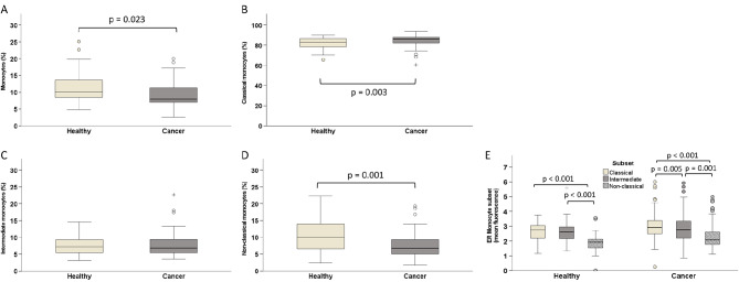
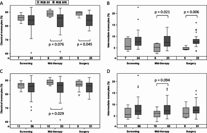
Results: When 40 healthy, age-matched women were compared with BC patients before treatment start, ER levels of monocytes did not differ, yet patients presented with a higher frequency of classical and fewer non-classical monocytes. Endocrine therapy triggered a significant increase in ER levels in all monocyte subsets, without affecting subset distribution. Vaccination had no overall impact on subset frequency and ER expression. Yet, a shift from intermediate to classical monocytes during therapy correlated with changes in plasma cytokines and chemokines and was significantly associated with low residual cancer burden in vaccinated patients. Without tecemotide, baseline ER levels in classical monocytes were significantly higher in women with good response to endocrine therapy.

Conclusions: This study identified classical monocytes to be associated with ER positive BC and with patient response to neoadjuvant endocrine treatment and cancer vaccination.

Keywords: Aromatase inhibitor; Breast cancer; Estrogen receptor; Letrozole; Monocyte; Stimuvax; Tecemotide.


Conflict of interest statement

The authors declare that they have no competing interests.

Figures

Fig. 1
Fig. 1
Schematic representation of the treatment schedule for BC patients receiving letrozole-based endocrine therapy with or without additional cyclophosphamide (CYC) administration and tecemotide vaccinations. Three blood draws (BD1-3) served to assess monocyte populations over the course of neoadjuvant therapy
Fig. 2
Fig. 2
Frequency of monocyte populations as determined by flow cytometry in peripheral blood of postmenopausal BC patients compared to healthy, age-matched women. (A) The percentage of all monocytes (among leukocytes) as well as the frequency of subsets within the monocyte population are shown by boxplots: (B) classical, (C) intermediate, (D) non-classical monocytes. (E) Comparison of ER expression levels between monocyte subsets as separately evaluated for BC patients and healthy women. Group differences are evaluated by non-parametric Mann-Whitney U (A-D) test or Wilcoxon signed-rank test (E). Circles indicate outliers with distances from the interquartile range (IQR) greater than 1.5 times the IQR, and crosses indicate extreme values with distances greater than 3 times the IQR
Fig. 3
Fig. 3
ER expression of total monocytes (A) and monocyte subsets (B) in the entire collective of 73 BC patients during neoadjuvant therapy as determined by flow cytometry at baseline (screening), after 12 weeks of letrozole administration (mid-therapy) and at the end of endocrine treatment (prior to surgery). Statistical analysis is based on Wilcoxon signed-rank test. Circles indicate outliers with distances from the interquartile range (IQR) greater than 1.5 times the IQR, and crosses indicate extreme values with distances greater than 3 times the IQR
Fig. 4
Fig. 4
Frequency of classical (A, C) and intermediate (B, D) monocytes in patients with RCB 0/I compared to RCB II/III, when analyzed for the tecemotide treatment arm (A, B) or the entire collective (C, D). Statistical analysis is based on Mann-Whitney U test. Circles and crosses indicate data points with distances from the interquartile range (IQR) greater than 1.5 times the IQR for RCB 0/I and RCB II/III responders, respectively
Fig. 5
Fig. 5
Analysis of matching breast cancer tissue retrieved by biopsy before neoadjuvant therapy (left panel) and surgically resected after treatment (right panel) from a patient with RCB 1 score and a rise in classical blood monocytes during neoadjuvant therapy. Whole tissue scans (A, B) and zoom-in regions (C-H) are shown of H&E stained sections (A, C, E) or immunofluorescence stainings (B, D, F) of CD14 (green), CD16 (red), ER (white) and DNA/cell nuclei (blue). For better resolution of monocyte subsets and ER expression, the total color overlay of image F is further split into CD14 (green), CD16 (red) in image G and ER (white) in image H. Pink arrows indicate CD14 + CD16 + monocytes (yellow color overlay of red and green), while pink asterisks identify classical CD14 + CD16- monocytes (green)
Fig. 6
Fig. 6
ER expression level of classical (A), intermediate (B) and non-classical (C) monocytes in patients with RCB 0/I compared to RCB II/III, when analyzed for the endocrine treatment arm without cancer vaccination. Group differences are evaluated by Mann-Whitney U test. Circles and crosses indicate data points with distances from the interquartile range (IQR) greater than 1.5 times the IQR for RCB 0/I and RCB II/III responders, respectively

References

    1. Sung H, Ferlay J, Siegel RL, Laversanne M, Soerjomataram I, Jemal A, Bray F. Global Cancer statistics 2020: GLOBOCAN estimates of incidence and Mortality Worldwide for 36 cancers in 185 countries. CA Cancer J Clin. 2021;71:209–49. - PubMed
    1. Benz CC. Impact of aging on the biology of breast cancer. Crit Rev Oncol Hematol. 2008;66:65–74. - PMC - PubMed
    1. Demicheli R, Ardoino I, Boracchi P, Coradini D, Agresti R, Ferraris C, Gennaro M, Hrushesky WJ, Biganzoli E. Recurrence and mortality according to estrogen receptor status for breast cancer patients undergoing conservative surgery. Ipsilateral breast tumour recurrence dynamics provides clues for tumour biology within the residual breast. BMC Cancer. 2010;10:656. - PMC - PubMed
    1. Fuentes N, Silveyra P. Estrogen receptor signaling mechanisms. Adv Protein Chem Struct Biol. 2019;116:135–70. - PMC - PubMed
    1. Burstein HJ. Systemic therapy for Estrogen Receptor-Positive, HER2-Negative breast Cancer. N Engl J Med. 2020;383:2557–70. - PubMed