. 2024 Oct 8;12(1):116.
doi: 10.1186/s40364-024-00658-x.

Single cell spatial profiling of FFPE splenic tissue from a humanized mouse model of HIV infection

Affiliations

Single cell spatial profiling of FFPE splenic tissue from a humanized mouse model of HIV infection

Guoxin Wu et al. Biomark Res. .

Abstract

Background: Latency remains a major obstacle to finding a cure for HIV despite the availability of antiretroviral therapy. Due to virus dormancy, limited biomarkers are available to identify latent HIV-infected cells. Profiling of individual HIV-infected cells is needed to explore potential latency biomarkers and to study the mechanisms of persistence that maintain the HIV reservoir.

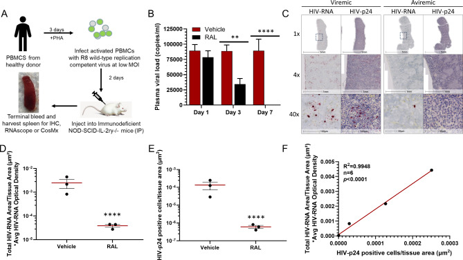
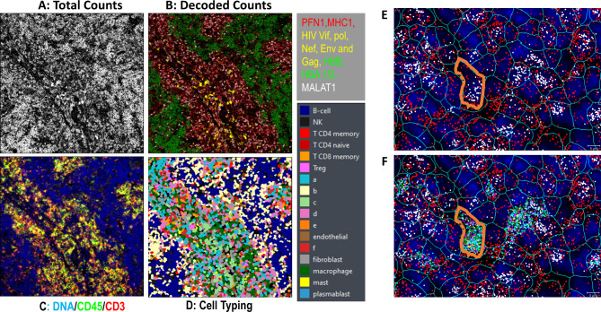
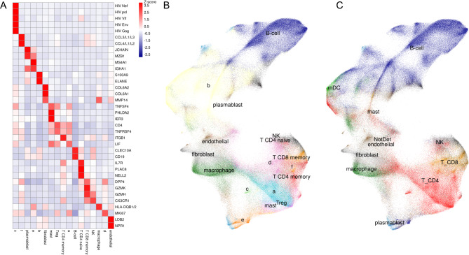
Methods: Single cell spatial transcriptomic characterization using the CosMx Spatial Molecular Imager platform was conducted to analyze HIV-infected cells in formalin-fixed paraffin-embedded sections of splenic tissue surgically obtained from an HIV-infected humanized mouse model. Regulation of over a thousand human genes was quantified in both viremic and aviremic specimens. In addition, in situ hybridization and immunohistochemistry were performed in parallel to identify HIV viral RNA- and p24-containing cells, respectively. Finally, initial findings from CosMx gene profiling were confirmed by isolating RNA from CD4 + T cells obtained from a person living with HIV on antiretroviral therapy following either PMA/Ionomycin or DMSO treatment. RNA was quantified using qPCR for a panel of targeted human host genes.

Results: Supervised cell typing revealed that most of the HIV-infected cells in the mouse spleen sections were differentiated CD4 + T cells. A significantly higher number of infected cells, 2781 (1.61%) in comparison to 112 (0.06%), and total HIV transcripts per infected cell were observed in viremic samples compared to aviremic samples, respectively, which was consistent with the data obtained from ISH and IHC. Notably, the expression of 55 genes was different in infected cells within tissue from aviremic animals compared to viremic. In particular, both spleen tyrosine kinase (SYK) and CXCL17, were expressed approximately 100-fold higher. This data was further evaluated against bulk RNA isolated from HIV-infected human primary CD4 + T cells. A nearly 6-fold higher expression of SYK mRNA was observed in DMSO-treated CD4 + T cells compared to those stimulated with PMA/Ionomycin.

Conclusion: This study found that the CosMx SMI platform is valuable for assessing HIV infection and providing insights into host biomarkers associated with HIV reservoirs. Higher relative expression of the SYK gene in aviremic-infected cells from the humanized mouse HIV model was consistent with levels found in CD4 + T cells of aviremic donors.

Keywords: Biomarker; CosMx SMI; FFPE; HIV; SYK; Single cell; Transcriptomic profiling; qPCR.


Conflict of interest statement

G.W., S.H.K., L.S., B.M., P.Z., C.J.B., J.M., C.C., T.R. and B.J.H. are current or former employees of Merck Sharp & Dohme Corp, a subsidiary of Merck & Co., Inc. Rahway, NJ, USA and may hold stock in Merck & Co., Inc. Rahway, NJ, USA. C.W., K.T., M.G. and L.P. are employees or former employees of NanoString Technologies, a Bruker Company declare no competing interests.

Figures

Fig. 1
Fig. 1
Characterization of humanized mouse HIV model: (A) Generation of HIV model in immunodeficient NOD-SCID-IL-2ry-/- mice with IP injection of HIV- infected human PBMC. (B) Plasma viral load (copies/ml) in vehicle and anti-viral integrase RAL-treated mice. (C) Imaging of viral RNA (RNAscope) and HIV p24 protein (IHC in FFPE spleen section) from HIV-infected vehicle treated (viremic) and anti-viral treated (aviremic) mice. (D) Quantitation of HIV viral RNA-containing cells in images from vehicle and RAL-treated mice. (E) Quantitation of HIV p24 protein-containing cells in tissues from vehicle and RAL-treated mice. (F) Correlation between tissue HIV viral RNA- and p24-containing cells in vehicle and anti-viral RAL-treated mice (n = 6)
Fig. 2
Fig. 2
CosMx SMI technology: (A) CosMx SMI raw data image representing total counts collected from 10 probes, (B) CosMx SMI raw data image representing decoded counts collected from 10 probes, (C) Image of CosMx morphology marker staining using anti-CD3 and anti-CD45 antibodies and DAPI, (D) CosMx cell typing result, (E) Individual cell cytoplasm and nucleus localization, and (F) HIV RNA located within an individual infected cell’s cytoplasm. Cell border is highlighted in orange
Fig. 3
Fig. 3
Gene Heatmap and UMAP cell clustering: (A) Genetic marker heatmap based on cell typing, (B) Semi-supervised cell clustering in UMAP, (C) Supervised UMAP cell clustering
Fig. 4
Fig. 4
Characterization of HIV-infected cells in viremic and aviremic conditions: (A) CosMx cell typing image of HIV-infected cells in viremic section, (B) CosMx cell typing image of HIV-infected cells in aviremic section, (C) Correlation between number of total infected cells and HIV transcripts per infected cell in viremic vs. aviremic sections, (D) Comparison of HIV-infected cell number based on the criteria of 10 or more normalized copies per cell and percentage of infected cells based on the total analyzed cells between viremic and aviremic sections
Fig. 5
Fig. 5
Comparison of 1000-panel human gene profiling between viremic and aviremic sections: (A) Gene expression of SYK and CXCL-17 were upregulated significantly (p < 0.0001) in infected vs. uninfected cells under aviremic conditions, (B) Upregulation of genes in infected vs. HIV-uninfected CD4 + T cells under viremic conditions (p < 0.05), (C) Upregulation of genes in HIV-infected CD4 + T cells in aviremic section as compared with that of viremic section (p < 0.05). Several genes including SYK and CXCL17 with p value < 0.0001, (D) Fold- change of CXCL17, SYK and S100A4 genes among infected vs. uninfected cells in aviremic vs. viremic conditions
Fig. 6
Fig. 6
Measurement of SYK gene in CD4 + T cells from aviremic donors: Measured HIV cell-associated RNA (A) and HIV p24 protein in culture supernatant (B) were significantly increased in PWH donor CD4 + T cells following PMA/Ionomycin stimulation. SYK mRNA level was significantly increased in aviremic CD4 + T cells as compared with that of T cells stimulated with PMA/Ionomycin for 48 h after the adjustment of either S100A4 gene (C) or CD4 gene (D)

References

    1. Kobayashi-Ishihara M, Tsunetsugu-Yokota Y. Post-transcriptional HIV-1 latency: a Promising Target for Therapy? Viruses. 2024;16:666. - PMC - PubMed
    1. Chou TC, Maggirwar NS, Marsden MD. HIV persistence, latency, and cure approaches: where are we now? Viruses. 2024;16:1163. - PMC - PubMed
    1. Broder S. The development of antiretroviral therapy and its impact on the HIV-1/AIDS pandemic. Antivir Res. 2010;85:1–18. - PMC - PubMed
    1. Einkauf KB, Osborn MR, Gao C, Sun W, Sun X, Lian X, et al. Parallel analysis of transcription, integration, and sequence of single HIV-1 proviruses. Cell. 2022;185:266–82. - PMC - PubMed
    1. Marcello A, Latronico T. Latency and reactivation of HIV-1. Microorganisms. 2020;8:76. - PubMed

LinkOut - more resources