Review
. 2025;103(6):521-545.
doi: 10.1159/000541881. Epub 2024 Oct 9.

The Immune Microenvironment in Prostate Cancer: A Comprehensive Review

Affiliations
Review

The Immune Microenvironment in Prostate Cancer: A Comprehensive Review

Rene Novysedlak et al. Oncology. 2025.

Abstract

Background: Prostate cancer (PCa) is a malignancy with significant immunosuppressive properties and limited immune activation. This immunosuppression is linked to reduced cytotoxic T cell activity, impaired antigen presentation, and elevated levels of immunosuppressive cytokines and immune checkpoint molecules. Studies demonstrate that cytotoxic CD8+ T cell infiltration correlates with improved survival, while increased regulatory T cells (Tregs) and tumor-associated macrophages (TAMs) are associated with worse outcomes and therapeutic resistance. Th1 cells are beneficial, whereas Th17 cells, producing interleukin-17 (IL-17), contribute to tumor progression. Tumor-associated neutrophils (TANs) and immune checkpoint molecules, such as PD-1/PD-L1 and T cell immunoglobulin-3 (TIM-3) are also linked to advanced stages of PCa. Chemotherapy holds promise in converting the "cold" tumor microenvironment (TME) to a "hot" one by depleting immunosuppressive cells and enhancing tumor immunogenicity.

Summary: This comprehensive review examines the immune microenvironment in PCa, focusing on the intricate interactions between immune and tumor cells in the TME. It highlights how TAMs, Tregs, cytotoxic T cells, and other immune cell types contribute to tumor progression or suppression and how PCa's low immunogenicity complicates immunotherapy.

Key messages: The infiltration of cytotoxic CD8+ T cells and Th1 cells correlates with better outcomes, while elevated T regs and TAMs promote tumor growth, metastasis, and resistance. TANs and natural killer (NK) cells exhibit dual roles, with higher NK cell levels linked to better prognoses. Immune checkpoint molecules like PD-1, PD-L1, and TIM-3 are associated with advanced disease. Chemotherapy can improve tumor immunogenicity by depleting T regs and myeloid-derived suppressor cells, offering therapeutic promise.

Keywords: CD4; CD8; Immunology; Immunotherapy; Metastatic; Myeloid-derived suppressor cells; Neutrophils; Prostate tumor; T cells; Tertiary lymphoid structures; Tumor-associated macrophages.


Conflict of interest statement

The Rene Novysedlak, Robin Bartolini, Lily Koumbas Foley, Miray Güney, Majd Al Khouri, Iva Benesova, Andrej Ozaniak, Vojtech Novak, Stepan Vesely, Pavel Pacas, Tomas Buchler, and Zuzana Ozaniak Strizova declare no conflict of interest. Tomas Buchler declares following research support: AstraZeneca, Roche, Bristol Myers Squibb, Exelixis, Merck KGaA, MSD, and Novartis; consulting fees from Bristol Myers Squibb, Astellas, Janssen, and Sanofi/Aventis; payment or honoraria for lectures, presentations, speakers’ bureaus, manuscript writing, or educational events from Ipsen, Bristol-Myers Squibb, AstraZeneca, Roche, Servier, Accord, MSD, and Pfizer. All unrelated to the present paper.

Figures

Fig. 1.
Fig. 1.
Representation of stromal cells infiltrating PCa. The figure illustrates the diverse stromal cell populations infiltrating PCa, encompassing macrophages, T cells, TANs, dendritic cells, MDSCs, and cancer-associated macrophages. These distinct cell types play pivotal roles in the TME, influencing tumor progression, immune response modulation, and therapeutic outcomes. Created with BioRender.com.
Fig. 2.
Fig. 2.
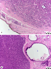
Histological images of PCa in patients with Gleason Score 3 (a) and Gleason Score 5 (b). The figure presents real histological sections from PCa patients, demonstrating different Gleason scores. a shows a Gleason score 3 tumor, where distinct features include: 1 – surgical margin stained with ink, 2 – fibrous capsule of the prostate, 3 – nerve, 4 – edge of the tumor (acinar adenocarcinoma of the prostate, Gleason score 3), and 5 – prostatic gland. In contrast, b highlights a more aggressive Gleason score 5 tumor, with key structures including: 1 – prostatic gland, and 2 – acinar adenocarcinoma of the prostate, Gleason score 4. These histological differences reflect varying degrees of tumor differentiation, which significantly impacts clinical prognosis and treatment planning.
Fig. 3.
Fig. 3.
The cancer immunity cycle. Diagram illustrating the sequential phases comprising the cancer immunity cycle, encompassing seven pivotal steps in generating an immune response against cancer. The process involves the release of tumor antigens from damaged cells, antigen presentation by DCs, activation of CD4 and CD8 T cells, followed by the orchestrated trafficking, infiltration, recognition, and eventual eradication of cancer cells via the cytotoxicity of CD8 T cells. Created with BioRender.com.
Fig. 4.
Fig. 4.
Roles of TAMs in PCa. TAMs participate in diverse functions including immune modulation, promotion of angiogenesis, extracellular matrix remodeling, fostering tumor growth, and facilitating metastasis. Their dynamic interactions influence tumor progression, therapeutic responses, and the overall TME milieu in PCa. Created with BioRender.com.
Fig. 5.
Fig. 5.
Contrasting mechanisms of trogoptosis and trogocytosis by tumor-associated neutrophils. Figure illustrating the distinct processes of trogoptosis and trogocytosis employed by TANs within the TME. Trogoptosis refers to the neutrophil-mediated induction of cancer cell death, while trogocytosis involves the transfer of cellular components or surface molecules from cancer cells to neutrophils, influencing immune responses, tumor progression, and microenvironmental interactions. Created with BioRender.com.
Fig. 6.
Fig. 6.
Differential signaling pathways of checkpoint receptors. The figure illustrates distinct signaling mechanisms initiated by checkpoint receptors, focusing on PD-1 and CTLA-4. The left segment demonstrates the interaction between programmed death-ligand 1 (PD-L1) and its receptor programmed cell death protein 1 (PD-1). The binding of PD-L1 to PD-1 results in the inhibition or blockade of T-cell receptor (TCR) intracellular signaling. This interaction leads to the attenuation of downstream signaling events crucial for T-cell activation, contributing to immunosuppressive effects within the cellular microenvironment. The right segment shows cytotoxic T-lymphocyte antigen-4 (CTLA-4) acting as a competitive receptor to CD28. Both CTLA-4 and CD28 compete for binding to the same ligands. CTLA-4 binding outcompetes CD28, resulting in a regulatory mechanism that dampens T-cell activation and reduces immune responses by inhibiting the essential costimulatory signals mediated by CD28. Created with BioRender.com.
Fig. 7.
Fig. 7.
Diversity of tertiary lymphoid organs (TLOs) in cancer. The figure illustrates the diverse cellular components, locations, and key variables associated with tertiary lymphoid organs (TLOs). Key cytokines and chemokines driving TLO formation and function are annotated. fDCs, follicular dendritic cells, FRCs, fibroblastic reticular cells. Created with BioRender.com.

References

    1. Stultz J, Fong L. How to turn up the heat on the cold immune microenvironment of metastatic prostate cancer. Prostate Cancer Prostatic Dis. 2021;24(3):697–717. - PMC - PubMed
    1. Sun BL. Immunotherapy in treatment of metastatic prostate cancer: an approach to circumvent immunosuppressive tumor microenvironment. Prostate. 2021;81(15):1125–34. - PubMed
    1. Apusiga K. Immune cell infiltration-based prognosis in prostate cancer: a review of current knowledge. Bull Natl Res Cent. 2023;47(1):131.
    1. Dong L, Myers KV, Pienta KJ. Understanding the tumor-immune microenvironment in prostate cancer. Curr Opin Oncol. 2021;33(3):231–7. - PubMed
    1. Liu J, Li Y, Yang D, Yang C, Mao L. Current state of biomarkers for the diagnosis and assessment of treatment efficacy of prostate cancer. Discov Med. 2019;27(150):235–43. - PubMed

MeSH terms