

Morphology of Roots and Canals of Maxillary First Premolars: A CBCT Study in a Peruvian Sample
- PMID: 39380790
- PMCID: PMC11461065
- DOI: 10.1155/2024/2341041
Morphology of Roots and Canals of Maxillary First Premolars: A CBCT Study in a Peruvian Sample
Abstract
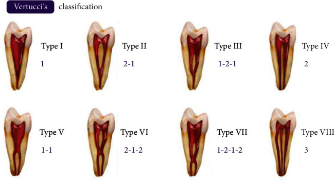
Introduction: It is important to take into account variations in structures related to dental pulp for planning the most adequate endodontic treatment management. The objective of this study was to determine the morphology of roots and canals of maxillary first premolars (MFPs) using cone-beam computed tomography (CBCT). Materials and Methods: This retrospective study included a sample of 392 CBCTs of Peruvian adults proportionally selected by sex, age, and quadrant. One MFP per individual was selected for evaluation by a calibrated evaluator based on the number of roots and canal configuration according to the Vertucci classification (VC; Cohen's κ ≥ 0.834). Pearson's χ 2 and Kruskal-Wallis tests were used with a significance level of P < 0.05. Results: Most MFP presented double roots (59.9%) and were VC type IV (52%). Morphology showed a nonsignificant difference by quadrants (P=0.994). A significant positive association was found between the presence of double roots and type IV in men and with older age, while single roots and type I and II were associated with women and younger age (P < 0.05). Double roots were associated with VC type IV (86%) and single roots with types III (34%), II (32%), and I (26%; P < 0.001). Conclusions: MFPs in a Peruvian sample presented a higher frequency of double roots with two separate canals. The morphology of root and canals was associated with age and sex.
Keywords: canal configuration; cone-beam computed tomography; maxillary premolars; root morphology.
Copyright © 2024 Juan Salvador Yanqui-Gómez et al.
Conflict of interest statement
The authors declare no conflicts of interest.
Figures


References
-
- Saber S. E. D. M., Ahmed M. H. M., Obeid M., Ahmed H. M. A. Root and Canal Morphology of Maxillary Premolar Teeth in an Egyptian Subpopulation Using Two Classification Systems: A Cone Beam Computed Tomography Study. International Endodontic Journal . 2019;52(3):267–278. doi: 10.1111/iej.13016. - DOI - PubMed
LinkOut - more resources
Full Text Sources
