. 2023 Sep 20:3:1184309.
doi: 10.3389/fnume.2023.1184309. eCollection 2023.

Radiolabeled iron oxide nanoparticles functionalized with PSMA/BN ligands for dual-targeting of prostate cancer

Affiliations

Radiolabeled iron oxide nanoparticles functionalized with PSMA/BN ligands for dual-targeting of prostate cancer

Danae Efremia Bajwa et al. Front Nucl Med. .

Abstract

Introduction: Prostate cancer (PCa) is the second most frequent cancer diagnosis in men and the fifth leading cause of death worldwide. Prostate Specific Membrane Antigen (PSMA) and Gastrin Releasing Peptide (GRP) receptors are overexpressed in PCa. In this study, we have developed iron oxide nanoparticles (IONs) functionalized with the Prostate Specific Membrane Antigen (PSMA) and Gastrin Releasing Peptide (GRP) ligands for dual targeting of Prostate cancer.

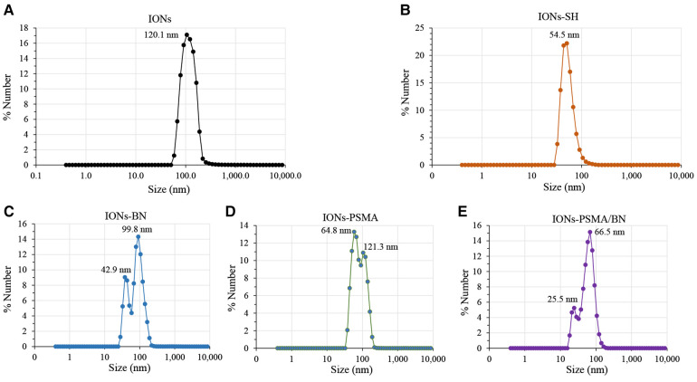
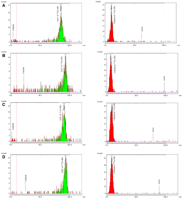
Methods: IONs were developed with a thin silica layer on their surface with MPTES (carrying -SH groups, IONs-SH), and they were coupled either with a pharmacophore targeting PSMA (IONs-PSMA) or with bombesin peptide (IONs-BN), targeting GRP receptors, or with both (IONs-PSMA/BN). The functionalized IONs were characterized for their size, zeta potential, and efficiency of functionalization using dynamic light scattering (DLS) and Fourier-Transform Infrared Spectroscopy (FT-IR). All the aforementioned types of IONs were radiolabeled directly with Technetium-99m (99mTc) and evaluated for their radiolabeling efficiency, stability, and binding ability on two different PCa cell lines (PC3 and LNCaP).

Results and discussion: The MTT assay demonstrated low toxicity of the IONs against PC3 and LNCaP cells, while the performed wound-healing assay further proved that these nanostructures did not affect cellular growth mechanisms. The observed hemolysis ratio after co-incubation with red blood cells was extremely low. Furthermore, the 99mTc-radiolabeled IONs showed good stability in human serum, DTPA, and histidine, and high specific binding rates in cancer cells, supporting their future utilization as potential diagnostic tools for PCa with Single Photon Emission Computed Tomography (SPECT) imaging.

Keywords: 99mTc; PSMA; SPECT; bispecific heterodimers; bombesin; iron oxide nanoparticles; prostate cancer.


Conflict of interest statement

The author EE declared that they were an editorial board member of Frontiers, at the time of submission. This had no impact on the peer review process and the final decision. The authors declare that the research was conducted in the absence of any commercial or financial relationships that could be construed as a potential conflict of interest.

Figures

Figure 1
Figure 1
Hydrodynamic diameter of IONs measured with DLS: (A) before silica gel thin layer modification (IONs); (B) after MPTES modification (IONs-SH); and (C–E) after functionalization with pharmacophores BN, PSMA and PSMA/BN.
Figure 2
Figure 2
Observed changes in ζp values of nanoparticles before (IONs) and after MPTES modification (IONs-SH) and after functionalization with PSMA (IONs-PSMA), BN (IONs-BN), or both PSMA and BN (IONs-PSMA/BN).
Figure 3
Figure 3
FT-IR structural characterization of compounds: (1) IONs, (2) IONs-SH, (3) IONs-BN (4) IONs-PSMA, (5) IONs-PSMA/BN, (6) PSMA, and (7) BN.
SCHEME 1
SCHEME 1
Chemical structures of the IONs functionalized with pharmacophores PSMA (IONs-PSMA), BN (IONs-BN), or both (IONs-PSMA/BN). The IONs were radiolabeled with 99mTc, either with its direct complexation to the IONs surface, using anhydrous SnCl2 as the reducing agent of Na[99mTc]TcO4, or by using the [99mTc][Tc(H2O)3(CO)3]+ precursor. The directly labeled IONs were further investigated due to their higher RCC.
Figure 4
Figure 4
ITLC-SG radiochemical analysis (mobile phase: sodium citrate 0.1 M) to determine colloids on the left and with acetone to determine free pertechnetate [99mTc]TcO4 on the right. The analysis refers to the direct complexation of 99mTc to the IONs surface for: (A) [99mTc]Tc-IONs-SH, (B) [99mTc]Tc-IONs-PSMA, (C) [99mTc]Tc-IONs-BN, and (D) [99mTc]Tc-IONs-PSMA/BN.
Figure 5
Figure 5
Results of hemolysis assay for (A) IONs-SH, (B) IONs-PSMA, (C) IONs-BN, and (D) IONs-PSMA/BN.
Figure 6
Figure 6
(A) Evaluation of cell viability via MTT assay in PC3 and (B) in LNCaP cells for IONs-SH, IONs-PSMA, IONs-BN, and IONs-PSMA/BN.
Figure 7
Figure 7
(A1,A2) Representative optical microscopy pictures of the scratch on PC3 and LNCaP cells (scale bar at 200 μm) and (B1,B2) quantified results of the wound closure (% WC). The scratch was monitored at different time points t = 0, 24, 48 and 72 h after incubation with IONs-SH, IONs-PSMA, IONs-BN, and IONs-PSMA/BN at C = 10 μgFe/ml.
Figure 8
Figure 8
Cell binding studies on LNCaP (A1–A4) and PC3 (B1–B4) cancer cells at 37°C and 4°C for the direct complexation of 99mTc to the IONs surface, [99mTc]Tc-labeled: IONs-SH (control), IONs-PSMA (LNCaP), IONs-BN (PC3) and IONs-PSMA/BN (LNCaP/PC3), where ACID represents the measured radioactivity in the acid wash (surface bound) and LYSATED the radioactivity measured after cells were lysed with NaOH (internalized). The total cell bound (=Surface + Internalized) radioactivity, with the statistical difference in comparison to IONs-SH (LNCaP: A3,A4, PC3: B3,B4), where (ns) p > 0.05, (*) p ≤ 0.05, (**) p ≤ 0.01, (***) p ≤ 0.001, (****) p ≤ 0.0001.

References

    1. Siegel RL, Miller KD, Jemal A. Cancer statistics, 2020. CA Cancer J Clin. (2020) 70(1):7–30. 10.3322/caac.21590 - DOI - PubMed
    1. Cornford P, Bellmunt J, Bolla M, Briers E, De Santis M, Gross T, et al. EAU-ESTRO-SIOG guidelines on prostate cancer. Part II: treatment of relapsing, metastatic, and castration-resistant prostate cancer. Eur Urol. (2017) 71(4):630–42. 10.1016/j.eururo.2016.08.002 - DOI - PubMed
    1. Mottet N, van den Bergh RCN, Briers E, Van den Broeck T, Cumberbatch MG, De Santis M, et al. EAU-EANM-ESTRO-ESUR-SIOG guidelines on prostate cancer—2020 update. Part 1: screening, diagnosis, and local treatment with curative intent. Eur Urol. (2021) 79(2):243–62. 10.1016/j.eururo.2020.09.042 - DOI - PubMed
    1. Bouchelouche K, Tagawa ST, Goldsmith SJ, Turkbey B, Capala J, Choyke P. PET/CT imaging and radioimmunotherapy of prostate cancer. Semin Nucl Med. (2011) 41(1):29–44. 10.1053/j.semnuclmed.2010.08.005 - DOI - PMC - PubMed
    1. De Rosales RTM. Potential clinical applications of bimodal PET-MRI or SPECT-MRI agents. J Label Compd Radiopharm. (2014) 57(4):298–303. - PMC - PubMed

LinkOut - more resources