. 2024 Sep 24:16:1459134.
doi: 10.3389/fnagi.2024.1459134. eCollection 2024.

NLRP3 inflammasome activation and pyroptosis are dispensable for tau pathology

Affiliations

NLRP3 inflammasome activation and pyroptosis are dispensable for tau pathology

Ine Paesmans et al. Front Aging Neurosci. .

Abstract

Background: Neuroinflammation is widely recognized as a key factor in the pathogenesis of Alzheimer's disease (AD), alongside ß-amyloid deposition and the formation of neurofibrillary tangles. The NLR family pyrin domain containing 3 (NLRP3) inflammasome, part of the innate immune system, has been implicated in the neuropathology of both preclinical amyloid and tau transgenic models. Activation of the NLRP3 pathway involves an initial priming step, which increases the expression of Nlrp3 and interleukin (IL)-1β, followed by the assembly of the NLRP3 inflammasome complex, comprising NLRP3, ASC, and caspase-1. This assembly leads to the proteolytic maturation of the pro-inflammatory cytokines IL-1β and IL-18. Additionally, the NLRP3 inflammasome induces Gasdermin D (GSDMD) cleavage, forming membrane pores through which IL-1β and IL-18 are secreted. Inhibition of NLRP3 has been shown to enhance plaque clearance by modulating microglial activation. Furthermore, blocking NLRP3 in tau transgenic mice has been found to reduce tau phosphorylation by affecting the activity of certain tau kinases and phosphatases.

Methods: In this study, organotypic brain slice cultures from P301S transgenic mice were treated with lipopolysaccharide (LPS) plus nigericin as a positive control or exposed to tau seeds (K18) to evaluate NLRP3 inflammasome activation. The effect of tau seeding on NLRP3 activity was further examined using Meso Scale Discovery (MSD) assays to measure IL1β secretion levels in the presence and absence of NLRP3 inhibitors. The role of NLRP3 activity was investigated in full-body Nlrp3 knockout mice crossbred with the tau transgenic P301S model. Additionally, full-body and microglia-selective Gsdmd knockout mice were crossbred with P301S mice, and tau pathology and neurodegeneration were evaluated at early and late stages of the disease using immunohistochemistry and biochemical assays.

Results: Activation of the NLRP3 pathway was observed in the mouse organotypic slice culture (OSC) model following stimulation with LPS and nigericin or exposure to tau seeds. However, Nlrp3 deficiency did not mitigate tauopathy or neurodegeneration in P301S mice in vivo, showing only a minor effect on plasma neurofilament (NF-L) levels. Consistently, Gsdmd deficiency did not alter tau pathology in P301S mice. Furthermore, neither full-body nor microglia-selective Gsdmd deletion had an impact on neuronal pathology or the release of pro-inflammatory cytokines.

Conclusion: The absence of key components of the NLRP3 inflammasome pathway did not yield a beneficial effect on tau pathology or neurodegeneration in the preclinical Tau-P301S mouse model of AD. Nonetheless, organotypic slice cultures could serve as a valuable ex vivo mechanistic model for evaluating NLRP3 pathway activation and pharmacological inhibitors.

Keywords: GSDMD; NLRP3; OSCs; P301S; neurodegeneration and tau pathology.


Conflict of interest statement

AB, DW, IP, KG, KV, and NO are employees of Jansssssen Pharmaceutica. The remaining authors declare that the research was conducted in the absence of any commercial or financial relationships that could be construed as a potential conflict of interest.

Figures

Figure 1
Figure 1
NLRP3 pathway activation in mouse organotypic slice cultures. (A) IL-1β measurement in the supernatant of OSCs stimulated with lipopolysaccharides (LPS) and nigericin and treated with NLRP3 small molecule inhibitor (MCC950 or Compound 1). Elevated IL-1β levels in LPS + nigericin condition, which are significantly reduced by increasing concentration of NLRP3 inhibitors. Figure represents two independent experiments combined to increase the replicates per condition. Quantitative data are shown as individual datapoint, mean with standard error. PBS and LPS n = 16, Nigericin: n = 10, 10 nM, Compound 1: 100 nM (n = 3), 300 nM (n = 3), 10 μM (n = 4); MCC950: 10 nM (n = 3), 300 nM (n = 4), 10 μM (n = 2). (B) OSCs supernatant shows elevated IL-1β levels after K18 seeding, which are significantly decreased by addition of 1 μM NLRP3 small molecule inhibitor called JNJ81977038. Data are shown as individual datapoint, mean with standard error. Significance ****p < 0.0001 by one-way ANOVA with multiple comparisons test.
Figure 2
Figure 2
No effect of Nlrp3 knockout on tau pathology by IHC. (A) AT100 immunohistochemistry demonstrated no significant effect on tau phosphorylation/aggregation in all analyzed regions of late-stage Nlrp3−/−xP301S vs. Nlrp3+/+xP301S mice. Brain stem, spinal cord, and midbrain showed a trend in reduced pathology for Nlrp3 deficiency, which was not significant. Early stage P301S mice displayed much lower aggregated tau levels compared to late-stage animals and no effect of Nlrp3 deletion on tauopathy was observed at this age in any of the regions analyzed. Image analysis data are shown as mean + individual values for each animal (average of two sections/animal). Early stage Nlrp3+/+xP301S: n = 24, Nlrp3−/−xP301S: n = 26; Late-stage Nlrp3+/+xP301S: n = 18, Nlrp3−/−xP301S: n = 19. No significance by unpaired t-test of age-matched groups. (B) Representative images of the AT00 IHC staining are demonstrated. Scale bars: 2.5 mm (overview images), 100 μm (higher magnification images). (C) Biochemical analysis of tau pathology in brainstem of Nlrp3xP301S mice. Aggregated tau levels were not significantly reduced in the sarcosyl-insoluble fraction or total homogenate of the brainstem of late-stage Nlrp3−/− vs. Nlrp3+/+xP301S mice. Early stage Nlrp3+/+xP301S: n = 23, Nlrp3−/−xP301S: n = 25; Late-stage Nlrp3+/+xP301S: n = 16, Nlrp3−/−xP301S: n = 16. Data are expressed as mean + individual values of each animal tested (values = average of two sections/mouse).
Figure 3
Figure 3
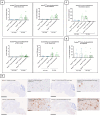
Microglia-specific Gsdmd targeting does not alter tau pathology by IHC or neurofilament levels in P301S mice. (A) No significant difference in AT100 levels is detected between GsdmdFL/FLxP301S mice and GsdmdFL/FLCx3cr1CreErt2xP301S mice, at early and late stage. Quantitative data are shown as mean + individual values for each animal. Early stage GsdmdFL/FLxP301S: n = ±6, GsdmdFL/FLCx3cr1CreErt2xP301S: n = ±4; Late-stage GsdmdFL/FLxP301S: n = ±10, GsdmdFL/FLCx3cr1CreErt2xP301S: n = ±12. No significance by unpaired t-test of age-matched groups. (B) Representative images of brain stem, midbrain, spinal cord gray matter, and cortex are shown. The whole brain sections are displayed at 2.5 mm, except for “GsdmdFL/FL Cx3cr1CreErt2xP301S late stage” at 5 mm and the spinal cord sections are displayed at 100 μm. (C) Late-stage P301S mice show higher levels of NF-L compared to early stage p301S mice and P301S heterozygous mice. However, no difference in plasma NF-L levels were detected between GsdmdFL/FL xP301S mice and GsdmdFL/FLCx3cr1CreErt2xP301S mice. Early stage GsdmdFL/FL xP301S n = 7, GsdmdFL/FLCx3cr1CreErt2xP301S: n = 5; Late-stage GsdmdFL/FL xP301S: n = 15, GsdmdFL/FLCx3sssscr1CreErt2xP301S: n = 14; GsdmdFL/FL xP301S HE: n = 6, GsdmdFL/FLCx3cr1CreErt2xP301S HE. No significance by unpaired t-test of age-matched groups. (D) No differences between GsdmdFL/FL xP301S and GsdmdFL/FLCx3cr1CreErt2xP301S mice on plasma IL-18 cytokine level measured with mesoscale. Early stage GsdmdFL/FL xP301S n = 6, GsdmdFL/FLCx3cr1CreErt2xP301S: n = 5; Late-stage GsdmdFL/FL xP301S: n = 11, GsdmdFL/FLCx3cr1CreErt2xP301S: n = 14. No significance by unpaired t-test of age-matched groups.
Figure 4
Figure 4
AT100 immunohistochemistry was not altered in Gsdmd deficient mice. (A) Immunohistochemistry with phospho-tau AT100 antibody showed no significant effect on tau pathology in GsdmdxP301S mice. No significant difference is detected between Nlrp3−/−xP301S vs. Nlrp3+/+xP301S mice at early and late stage. Quantitative data are shown as mean + individual values for each animal. Early stage Nlrp3+/+xP301S: n = ±10, Nlrp3−/−xP301S: n = ±15; Late-stage Nlrp3+/+xP301S: n = ±25, Nlrp3−/−xP301S: n = ±20. No significance by unpaired t-test of age-matched groups. (B) Representative images of brain stem, midbrain, spinal cord, and cortex are shown. The whole brain sections are displayed at 2.5 mm and the spinal cord sections are displayed at 100 μm.
Figure 5
Figure 5
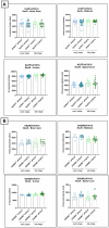
Late-stage Nlrp3 effect on neurofilament levels and no effect of Gsdmd in P301S mice. (A) Neurofilament light (NF-L) levels were quantified in plasma of Nlrp3xP301S mice as a surrogate marker for neurodegeneration in the CNS. At late stage, NF-L levels were significantly reduced in Nlrp3 knockout mice, compared to age-related Nlrp3 WT mice. No effect was demonstrated in early stage P301S mice. Early stage Nlrp3+/+xP301S: n = 23, Nlrp3−/−xP301S: n = 27; late-stage Nlrp3+/+xP301S: n = 18, Nlrp3−/−xP301S: n = 19. **p < 0.01 by paired t-test of age-matched groups. (B) No effect of GSDMD on plasma NF-L levels were observed in the tau transgenic P301S mouse model. WT: n = 17; Early stage Nlrp3+/+xP301S: n = 9, Nlrp3−/−xP301S: n = 21; Late-stage Nlrp3+/+xP301S: n = 27, Nlrp3−/−xP301S: n = 19. No significance by unpaired t-test of age-matched groups.
Figure 6
Figure 6
No effect of Nlrp3 or Gsdmd deletion on neuronal count in P301S mice. (A) The number of neurons was calculated in brain regions of Nlrp3xP301S mice at early and late stage. No effect of Nlrp3 on NeuN levels was demonstrated in the different stages nor the different brain regions of P301S mice. Early stage Nlrp3+/+xP301S: n = ±24, Nlrp3−/−xP301S: n = ±26; late-stage Nlrp3+/+xP301S: n = 18, Nlrp3−/−xP301S: n = ±19. No significance by unpaired t-test of age-matched groups. (B) Immunohistochemistry with NeuN antibody was also performed on early and late-stage Gsdmd−/−xP301S vs. Gsdmd+/+xP301S, demonstrating no effect of GSDMD. Early stage Gsdmd+/+xP301S: n = 10, Gsdmd−/−xP301S: n = ±12; Late-stage Gsdmd+/+xP301S: n = ±20, Gsdmd−/−xP301S: n = ±15. No significance by unpaired t-test of age-matched groups.
Figure 7
Figure 7
No impact of Gsdmd and Nlrp3 deficiency on plasma IL-18 levels in P301S mice. (A) Mesoscale measurement of pro-inflammatory IL-18 levels in plasma of P301S mice. No significant difference between Nlrp3 knockout mice vs. Nlrp3 WT mice in early and late-stage P301S mice. Early stage Nlrp3+/+xP301S: n = 18, Nlrp3−/−xP301S n = 26; Late-stage Nlrp3+/+xP301S: n = 16, Nlrp3−/−xP301S: n = 18. No significance by paired t-test of age-matched groups. (B) IL-18 concentrations were additionally quantified in plasma of early and late-stage Gsdmd−/−xP301S vs. Gsdmd+/+xP301S, demonstrating no effect of GSDMD. WT: n = 15; Early stage Nlrp3+/+xP301S: n = 8, Nlrp3−/−xP301S: n = 21; Late-stage Nlrp3+/+xP301S: n = 26, Nlrp3−/−xP301S: n = 19. No significance by paired t-test of age-matched groups.

References

    1. Allen B., Ingram E., Takao M., Smith M. J., Jakes R., Virdee K., et al. . (2002). Abundant tau filaments and nonapoptotic neurodegeneration in transgenic mice expressing human P301S tau protein. J. Neurosci. 22, 9340–9351. doi: 10.1523/JNEUROSCI.22-21-09340.2002, PMID: - DOI - PMC - PubMed
    1. Arends Y. M., Duyckaerts C., Rozemuller J. M., Eikelenboom P., Hauw J. J. (2000). Microglia, amyloid and dementia in Alzheimer disease. Neurobiol. Aging 21, 39–47. doi: 10.1016/S0197-4580(00)00094-4, PMID: - DOI - PubMed
    1. Bacioglu M., Maia L. F., Preische O., Schelle J., Apel A., Kaeser S. A., et al. . (2016). Neurofilament light chain in blood and CSF as marker of disease progression in mouse models and in neurodegenerative diseases. Neuron 91, 56–66. doi: 10.1016/j.neuron.2016.05.018 - DOI - PubMed
    1. Bamberger M. E., Harris M., McDonald D., Husemann J., Landreth G. (2003). A cell surface receptor complex for fibrillar beta-amyloid mediates microglial activation. J. Neurosci. 23, 2665–2674. doi: 10.1523/JNEUROSCI.23-07-02665.2003, PMID: - DOI - PMC - PubMed
    1. Beach T. G., Walker R., McGeer E. G. (1989). Patterns of gliosis in alzheimer’s disease and aging cerebrum. Glia 2, 420–436. doi: 10.1002/glia.440020605 - DOI - PubMed

LinkOut - more resources