. 2024 Sep 24:15:1325286.
doi: 10.3389/fendo.2024.1325286. eCollection 2024.

ATF4-dependent and independent mitokine secretion from OPA1 deficient skeletal muscle in mice is sexually dimorphic

Affiliations

ATF4-dependent and independent mitokine secretion from OPA1 deficient skeletal muscle in mice is sexually dimorphic

Jennifer Streeter et al. Front Endocrinol (Lausanne). .

Abstract

Introduction: Reducing Optic Atrophy 1 (OPA1) expression in skeletal muscle in male mice induces Activation Transcription Factor 4 (ATF4) and the integrated stress response (ISR). Additionally, skeletal muscle secretion of Fibroblast Growth Factor 21 (FGF21) is increased, which mediates metabolic adaptations including resistance to diet-induced obesity (DIO) and glucose intolerance in these mice. Although FGF21 induction in this model can be reversed with pharmacological attenuation of ER stress, it remains to be determined if ATF4 is responsible for FGF21 induction and its metabolic benefits in this model.

Methods: We generated mice with homozygous floxed Opa1 and Atf4 alleles and a tamoxifen-inducible Cre transgene controlled by the human skeletal actin promoter to enable simultaneous depletion of OPA1 and ATF4 in skeletal muscle (mAO DKO). Mice were fed high fat (HFD) or control diet and evaluated for ISR activation, body mass, fat mass, glucose tolerance, insulin tolerance and circulating concentrations of FGF21 and growth differentiation factor 15 (GDF15).

Results: In mAO DKO mice, ATF4 induction is absent. Other indices of ISR activation, including XBP1s, ATF6, and CHOP were induced in mAO DKO males, but not in mOPA1 or mAO DKO females. Resistance to diet-induced obesity was not reversed in mAO DKO mice of both sexes. Circulating FGF21 and GDF15 illustrated sexually dimorphic patterns. Loss of OPA1 in skeletal muscle increases circulating FGF21 in mOPA1 males, but not in mOPA1 females. Additional loss of ATF4 decreased circulating FGF21 in mAO DKO male mice, but increased circulating FGF21 in female mAO DKO mice. Conversely, circulating GDF15 was increased in mAO DKO males and mOPA1 females, but not in mAO DKO females.

Conclusion: Sex differences exist in the transcriptional outputs of the ISR following OPA deletion in skeletal muscle. Deletion of ATF4 in male and female OPA1 KO mice does not reverse the resistance to DIO. Induction of circulating FGF21 is ATF4 dependent in males, whereas induction of circulating GDF15 is ATF4 dependent in females. Elevated GDF15 in males and FGF21 in females could reflect activation by other transcriptional outputs of the ISR, that maintain mitokine-dependent metabolic protection in an ATF4-independent manner.

Keywords: FGF21; insulin resistance; integrated stress response; mitochondria; obesity.


Conflict of interest statement

The authors declare that the research was conducted in the absence of any commercial or financial relationships that could be construed as a potential conflict of interest.

Figures

Figure 1
Figure 1
Quantification of OPA1 and ATF4 Expression in mOPA1 KO and mAO DKO mice. WT mOPA1 males (dark orange), mOPA1 KO males (light orange), WT mAO males (dark green), mAO DKO males (light green), WT MOPA1 females (dark yellow), mOPAI KO females (light yellow), WT MAO females (dark purple), and mAO DKO females (light purple). Representative immunoblots of OPA1 protein in gastrocnemius (GASTROC) muscle from 22-week-old male (A) and female (B) WT, mOPA1 knock out (KO), and mOPA1/mATF4 double KO (DKO) mice and densitometric analysis of OPA1 protein normalized by GAPDH in OPA1 males (C, n=5-8), AO males (D, n= 5-7), OPA1 females (E, n=11), and AO females (F, n=10-11). OPA1 mRNA quantification in GASTROC muscle from 22-week old males (G, n=9-12) and females (H, n=6-14), fold change vs OPA1 WT. ATF4 mRNA quantification in gastrocnemius muscle from 22-week old males (I, n=10-14) and females (J, n=9-14), fold change vs OPA1 WT. OPA1 (mOPA1 mice), AO (mATF4 OPA1 DKO mice), WT (wildtype), KO (knock out), DKO (double knockout), F (female), M (male). ROUT method was used to identify and remove outliers, Q=10%. Created using BioRender.com.
Figure 2
Figure 2
ER Stress Markers mRNA Expression: mRNA quantification in gastrocnemius (gastroc) muscle from 22-week old mOPA1 males and mAO males (A-D), mOPA1 females and mAO females (E-H) of XBP1 S/T (spliced/total, C and G), ATF6 (A, E), BiP (B, F), and CHOP (D, H), fold change vs OPA1 WT (n=4-11). Created using BioRender.com.
Figure 3
Figure 3
Body Mass and Fat Mass. Body mass (A–D), body fat mass (E–H) and body fat percent (I–L) after 10 weeks of high fat diet (HFD, circles) or control diet (CD, triangles). P values determined through Brown-Forsythe and Welch one-way ANOVA with Dunnett’s T3 multiple comparisons test. Comparison pairs shown here: mOPA1 or mATF4 OPA1 (AO) wildtype (WT) males (M) or females (F) on CD vs HFD, WT on HFD vs mOPA1 knockout (KO) or m ATF4 OPA1 double KO (AO DKO) on HFD, and OPA1 KO or AO DKO on CD vs HFD. Created using BioRender.com.
Figure 4
Figure 4
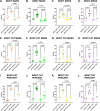
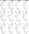
Loss of muscle mass in mOPA1 and mAO DKO mice. mOPAI KO males have a loss in lean muscle mass in multiple muscle groups including gastrocnemius (GASTROC), soleus (SOL), and tibialis anterior (TA) (A, E, I). mOPA1 females have decreased muscle mass in all muscle groups (C, G, K). MAO DKO males and females have significant loss of muscle mass compared to WT (B, D, F, H, J, L). Created using BioRender.com.
Figure 5
Figure 5
Glucose Tolerance Testing (GTT) and Insulin Tolerance Testing (ITT). GTT and Area under the curve (AUC) for males (A-C, n=3-7), and females (D-F, n=5-12), male ITT (G-I, n=3-6), and female ITT (J-L, n=3-6). Created using BioRender.com.
Figure 6
Figure 6
FGF21 and GDF15 levels in serum and skeletal muscle. Serum FGF21 (A males n=8-12, B females n=5- 9). Serum GDF15 (C males n-5-8, D females n-4-8). Tissue source of FGF21 and GDF15 assessed by measurement of mRNA induction in skeletal muscle (n=4-11): soleus (SOL, E-H), tibialis anterior (TA, I-L), and gastrocnemius (GAS, M-P). Created using BioRender.com.
Figure 7
Figure 7
FGF21 and GDF15 levels in liver and fat. Tissue source of FGF21 and GDF15 assessed by measurement of mRNA induction in liver (A-D, n=6-10), brown adipose tissue (BAT, E-H, n=4-8), and inguinal fat (IF, I-L, n-5-8). Created using BioRender.com.

References

    1. Neill G, Masson GR. A stay of execution: ATF4 regulation and potential outcomes for the integrated stress response. Front Mol Neurosci. (2023) 16:1112253. doi: 10.3389/fnmol.2023.1112253 - DOI - PMC - PubMed
    1. Rzymski T, Harris AL. The unfolded protein response and integrated stress response to anoxia. Clin Cancer Res. (2007) 13(9):2537–40. doi: 10.1158/1078-0432.CCR-06-2126 - DOI - PubMed
    1. Uebanso T, Taketani Y, Yamamoto H, Amo K, Ominami H, Arai H, et al. . Paradoxical regulation of human FGF21 by both fasting and feeding signals: is FGF21 a nutritional adaptation factor? PloS One. (2011) 6(9):e22976. doi: 10.1371/journal.pone.0022976 - DOI - PMC - PubMed
    1. Fisher FM, Kim M, Doridot L, Cunniff JC, Parker TS, Levine DM, et al. . A critical role for ChREBP-mediated FGF21 secretion in hepatic fructose metabolism. Mol Metab. (2017) 6(1):14–21. doi: 10.1016/j.molmet.2016.11.008 - DOI - PMC - PubMed
    1. Nishimura T, Nakatake Y, Konishi M, Itoh N. Identification of a novel FGF, FGF-21, preferentially expressed in the liver. Biochim Biophys Acta. (2000) 1492(1):203–6. doi: 10.1016/S0167-4781(00)00067-1 - DOI - PubMed

MeSH terms

LinkOut - more resources