. 2024 Sep 13;27(10):110953.
doi: 10.1016/j.isci.2024.110953. eCollection 2024 Oct 18.

Platelet factors ameliorate thoracic aortic aneurysm and dissection by inhibiting the FGF-FGFR cascade activation in aortic-endothelial cell

Affiliations

Platelet factors ameliorate thoracic aortic aneurysm and dissection by inhibiting the FGF-FGFR cascade activation in aortic-endothelial cell

Jizhong Wang et al. iScience. .

Abstract

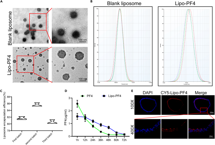
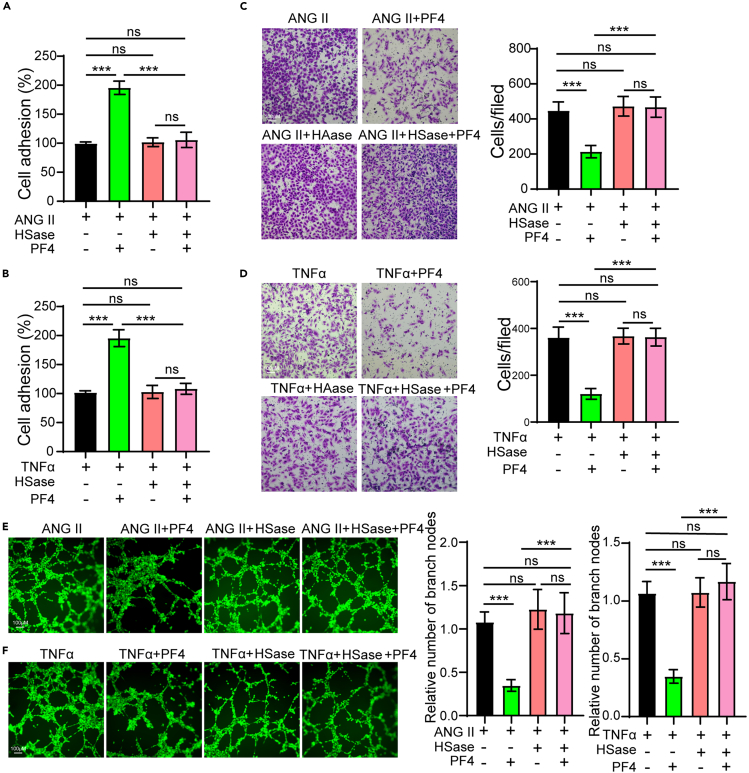
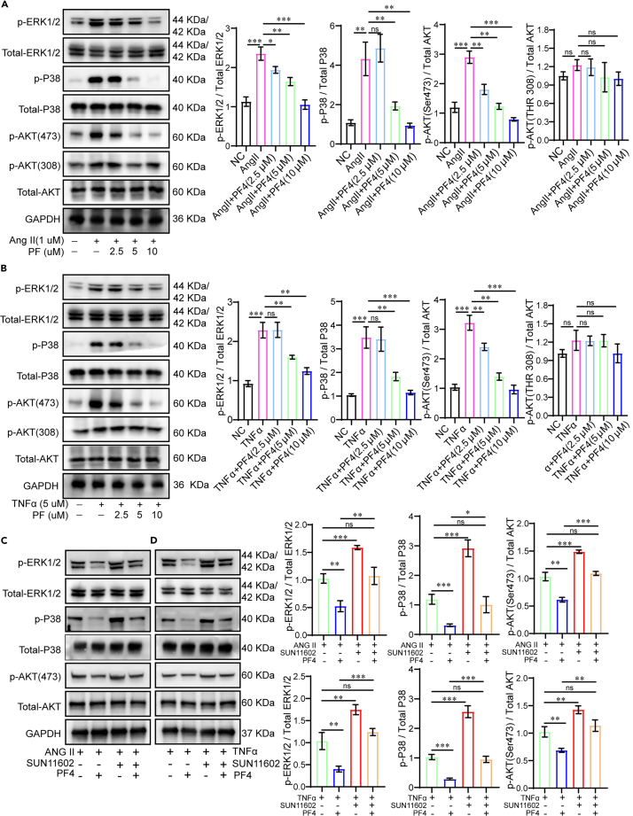
Thoracic aortic aneurysm and dissection (TAAD) is closely associated with vascular endothelial dysfunction. Platelet factor 4 (PF4) is crucial for maintaining vascular endothelial cell homeostasis. However, whether PF4 can influence the progression of TAAD remains unknown. In the present study, we constructed a liposome-encapsulated PF4 nanomedicine and verified its effect on BAPN-induced TAAD in vivo. We found that liposome PF4 nanoparticles (Lipo-PF4), more effectively than PF4 alone, inhibited the formation of TAAD. In vitro, PF4 improved endothelial cell function under pathological conditions by inhibiting migratory and angiogenic abilities of human aortic endothelial cells (HAECs). Mechanically, PF4 inhibited the development of TAAD and improved HAECs function by combining with heparin sulfate and blocking fibroblast growth factor-fibroblast growth factor receptor (FGF-FGFR) signaling. Taken together, we developed a nano-drug (Lipo-PF4) that effectively ameliorates the progression of TAAD by improving endothelial function. Lipo-PF4 is expected to be a therapeutic option for TAAD in the future.

Keywords: Biological sciences; Cardiovascular medicine; Drug delivery system; Therapeutics.


Conflict of interest statement

The authors declare no competing interests.

Figures

None
Graphical abstract
Figure 1
Figure 1
PF4 improves endothelial cell function in pathological conditions (A) Human aortic endothelial cells were treated with 1 μM AngII alone, 1 μM AngII plus PF4 at the indicated concentrations. Cell adhesion was measured 48 h later using CCK8 assay. (B) Human aortic endothelial cells were treated with 5 μM TNFα alone, 5 μM TNFα plus PF4 at the indicated concentrations. Cell adhesion was measured 48 h later using CCK8 assay. (C and D) Human aortic endothelial cells were treated with 1 μM AngII or 5 μM TNFα alone, AngII or TNFα plus PF4 at the indicated concentrations for 36 h, treated cells were seeded onto the upper chamber for 12 h, cells on the lower side of the filter were detected using an inverted microscope. (E and F) Human aortic endothelial cells were treated with 1 μM AngII or 5 μM TNFα alone, AngII or TNFα plus PF4 at the indicated concentrations for 36 h, treated cells were seeded onto the matrigel for 4 h in a 24-well plate. After incubating with 2 μM calcein AM for 30 min, invert the fluorescence microscope for detection. Data are expressed as mean ± SD. ∗p < 0.05, ∗ ∗p < 0.01, ∗ ∗ ∗p < 0.001.
Figure 2
Figure 2
Liposome encapsulation treatment reduces the degradation rate of PF4 protein in mice (A) PF4-SOD liposome was made through the rotation-evaporation method. Its morphology and diameter were detected using an electronic microscope. (B) Detection of liposome particle size using dynamic light scattering method. (C) ELISA assay for detecting the encapsulation efficiency of liposomes. (D) 100 μg/kg PF4 or PF4 liposome was injected into 8-week-old C57 mice through the tail vein, and the concentration of PF4 in the mouse blood was detected using ELISA assay at different time points. (E) After 100 μg/kg PF4 liposome-CY5 was injected into 8-week-old C57 mice for 12 h through the tail vein, the PF4 liposome-CY5 distribution was detected by immunofluorescence. Data are expressed as mean ± SD. ∗p < 0.05, ∗ ∗p < 0.01, ∗ ∗ ∗p < 0.001.
Figure 3
Figure 3
Liposome-PF4 nanoparticles repress thoracic aortic dissection formation by improving endothelial function (A) Mice were treated with liposome-PF4, PF4, or empty liposome once every three days at 4 weeks. Three days after the first administration, mice were treated with 0.25% BAPN (β-aminopropionitrile monofumarate) for 21 days. Subsequently, mice were treated with 1 μg/kg per minute AngII using minipump (n = 10, per group). (B and C) Representative images of mice aorta were shown. (D and E) maximum aortic diameter was measured (n = 10, per group). (F) AD incidence was statistically analyzed. (G) Representative images of mouse aorta stained with CD31 (red) and DAPI (blue). (H) Effect of PF4 on MMP activities in cultured supernatant of vascular wall endothelial cells by gelatin zymography. (I) Representative macroscopic images of aorta sections were stained with hematoxylin and eosin (H&E), Masson, and elastic-Van Gieson (EVG) staining. (J) Quantification of elastin integrity in each group of mouse aortas. Data are expressed as mean ± SD. ∗p < 0.05, ∗ ∗p < 0.01, ∗ ∗ ∗p < 0.001.
Figure 4
Figure 4
PF4 improves endothelial cell function by combining with heparin sulfate (A and B) After incubated with HSase (1 mU/ml, Sigma-Aldrich) for 4 h, cells were treated with 1 μM AngII or 5 μM TNFα alone, AngII or TNFα plus PF4 (10 μM) for 48 h, Cell adhesion was measured using CCK8 assay. (C and D) After incubated with HSase (1 mU/ml, Sigma-Aldrich) for 4 h, cells were treated with 1 μM AngII or 5 μM TNFα alone, AngII or TNFα plus PF4 (10 μM) for 36 h, treated cells were seeded onto the upper chamber for 12 h, cells on the lower side of the filter were detected using an inverted microscope. (E and F) After incubated with HSase (1 mU/ml, Sigma-Aldrich) for 4 h, cells were treated with 1 μM AngII or 5 μM TNFα alone, AngII or TNFα plus PF4(10 μM) for 36 h, treated cells were seeded onto the matrigel for 4 h in a 24-well plate. After incubating with 2 μM calcein AM for 30 min, the vascular cyclization ability of cells is detected. Data are expressed as mean ± SD. ∗p < 0.05, ∗ ∗p < 0.01, ∗ ∗ ∗p < 0.001.
Figure 5
Figure 5
PF4 represses thoracic aortic dissection formation and improves endothelial cell function by inhibiting the activation of FGF-FGFR (A) Differential gene analysis between BAPN group and Lipo-PF4 group. Mice were treated with liposome-PF4 once every three days at 3 weeks. Three days after the first administration, mice were treated with 0.25% BAPN (β-aminopropionitrile monofumarate) for 14 days (n = 9, per group).Mouse aortic intima were isolated, and the aortic intima of three mice were mixed into one sample for transcriptome sequencing, and performed differential gene analysis. N: BAPN group; P: lipo-PF4+BAPN group. (B) GO analysis of differential genes. (C) Mice were treated with liposome-PF4 or liposome-PF4 plus SUN11602 once every three days at 3 weeks. Three days after the first administration, mice were treated with 0.25% BAPN (β-aminopropionitrile mono fumarate) for 25 days (n = 10, per group). (D) Representative images of mice aorta were shown. (E) Maximum aortic diameter was measured (n = 10, per group). (F) AD incidence was statistically analyzed. (G) Effect of PF4 and SUN11602 on MMP activities in cultured supernatant of vascular wall endothelial cells by gelatin zymography. (H) Representative macroscopic images of aorta sections were stained with hematoxylin and eosin (H&E), Masson, and elastic-Van Gieson (EVG) staining. (I) Quantification of elastin integrity in each group of mouse aortas. (J) Representative images of mouse aorta stained with CD31 (red) and DAPI (blue). Data are expressed as mean ± SD. ∗p < 0.05, ∗ ∗p < 0.01, ∗ ∗ ∗p < 0.001.
Figure 6
Figure 6
PF4 represses MAPK signal transduction by blocking FGFs-FGFR (A and B) Human aortic endothelial cells were treated with 1 μM AngII or 5 μM TNFα alone, 1 μM AngII or 5 μM TNFα plus PF4 at the indicated concentrations for 24 h. The phosphorylated and total levels of Erk1/2, P38, Akt (Ser 473), and Akt (Thr 308) were detected by western blotting analysis (n = 3). (C and D) Cells were treated with 1 μM AngII or 5 μM TNFα alone, AngII or TNFα plus PF4(10 μM) and SUN11602(10 μM) for 24 h. The phosphorylated and total levels of Erk1/2, P38, and Akt (Ser 473) were detected by western blotting analysis (n = 3). Density ratios of phosphorylated proteins to total proteins were presented as the mean ± standard deviation. ∗p < 0.05, ∗ ∗p < 0.01, ∗ ∗ ∗p < 0.001.

References

    1. Pinard A., Jones G.T., Milewicz D.M. Genetics of Thoracic and Abdominal Aortic Diseases. Circ. Res. 2019;124:588–606. doi: 10.1161/circresaha.118.312436. - DOI - PMC - PubMed
    1. Famularo M., Meyermann K., Lombardi J.V. Aneurysmal degeneration of type B aortic dissections after thoracic endovascular aortic repair: A systematic review. J. Vasc. Surg. 2017;66:924–930. doi: 10.1016/j.jvs.2017.06.067. - DOI - PubMed
    1. Isselbacher E.M. Thoracic and abdominal aortic aneurysms. Circulation. 2005;111:816–828. doi: 10.1161/01.Cir.0000154569.08857.7a. - DOI - PubMed
    1. Halushka M.K., Angelini A., Bartoloni G., Basso C., Batoroeva L., Bruneval P., Buja L.M., Butany J., d'Amati G., Fallon J.T., et al. Consensus statement on surgical pathology of the aorta from the Society for Cardiovascular Pathology and the Association For European Cardiovascular Pathology: II. Noninflammatory degenerative diseases - nomenclature and diagnostic criteria. Cardiovasc. Pathol. 2016;25:247–257. doi: 10.1016/j.carpath.2016.03.002. - DOI - PubMed
    1. Cai Z., Yarovoi S.V., Zhu Z., Rauova L., Hayes V., Lebedeva T., Liu Q., Poncz M., Arepally G., Cines D.B., Greene M.I. Atomic description of the immune complex involved in heparin-induced thrombocytopenia. Nat. Commun. 2015;6:8277. doi: 10.1038/ncomms9277. - DOI - PMC - PubMed

LinkOut - more resources