. 2025 Jan 2;24(1):118-130.
doi: 10.1158/1535-7163.MCT-24-0163.

Tumor Integrin-Targeted Glucose Oxidase Enzyme Promotes ROS-Mediated Cell Death that Combines with Interferon Alpha Therapy for Tumor Control

Affiliations

Tumor Integrin-Targeted Glucose Oxidase Enzyme Promotes ROS-Mediated Cell Death that Combines with Interferon Alpha Therapy for Tumor Control

Jordan A Stinson et al. Mol Cancer Ther. .

Abstract

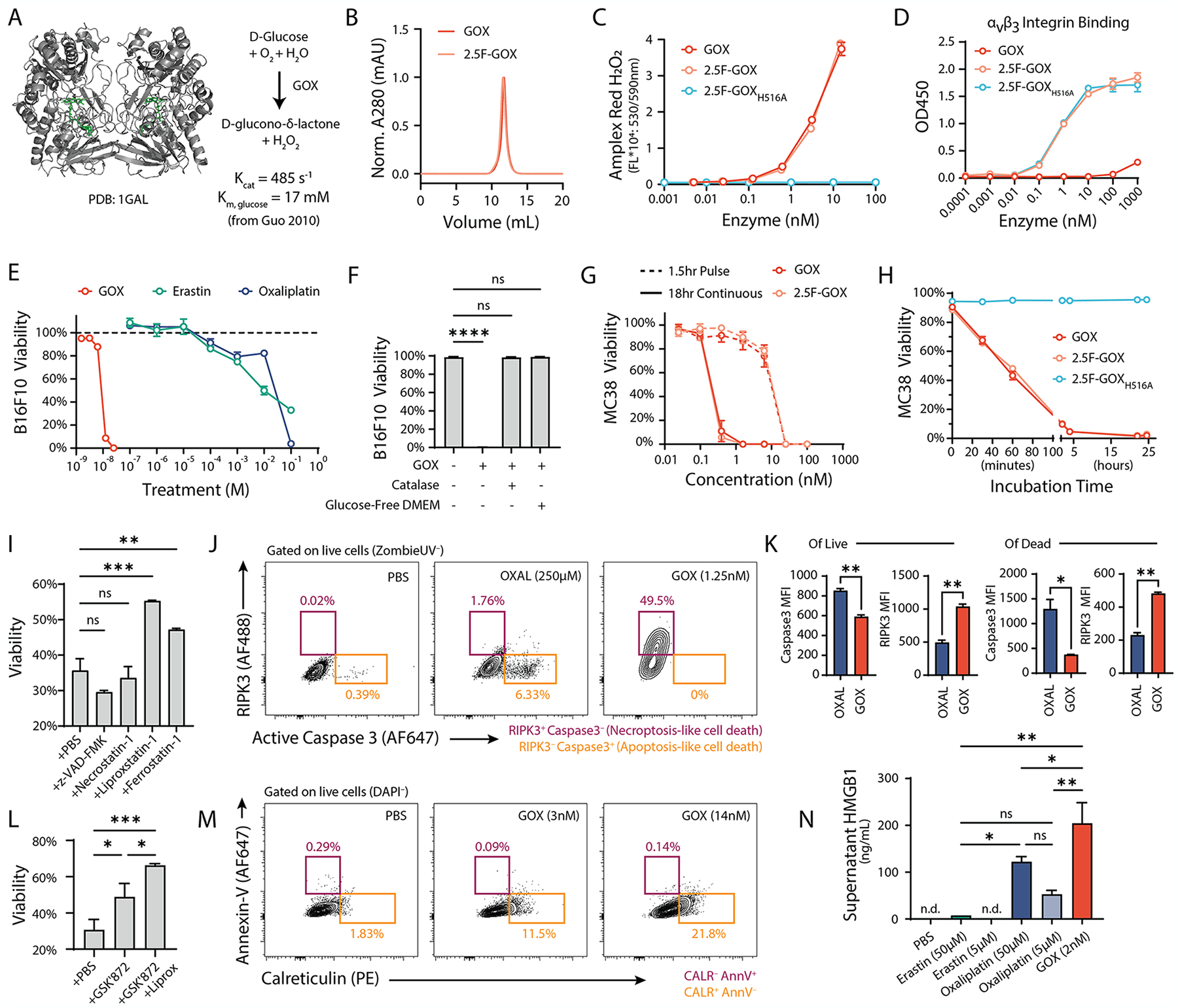
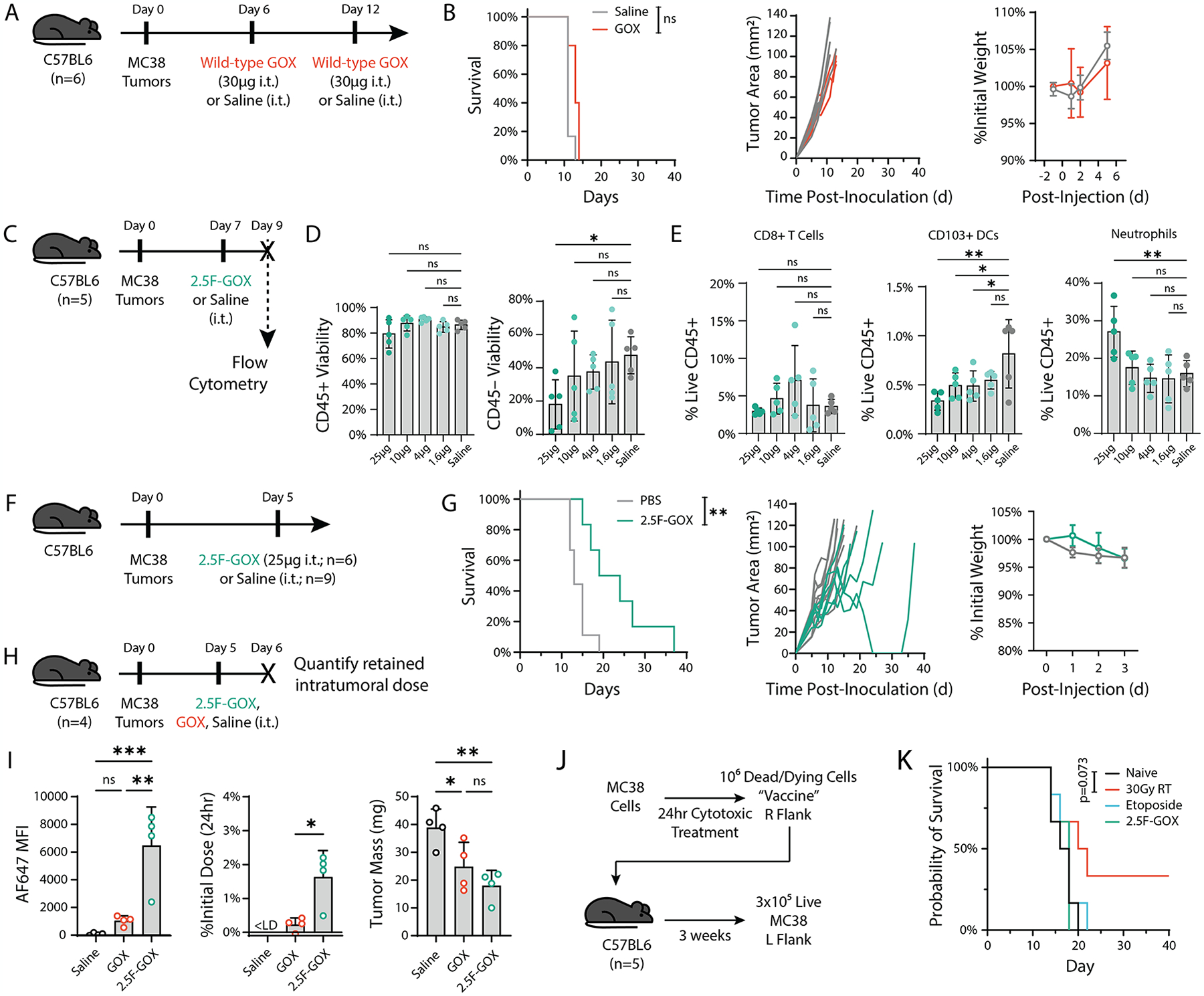
Although heightened intratumoral levels of reactive oxygen species (ROS) are typically associated with a suppressive tumor microenvironment, under certain conditions ROS contribute to tumor elimination. Treatment approaches, including some chemotherapy and radiation protocols, increase cancer cell ROS levels that influence their mechanism of cell death and subsequent recognition by the immune system. Furthermore, activated myeloid cells rapidly generate ROS upon encounter with pathogens or infected cells to eliminate disease, and recently, this effector function has been noted in cancer contexts as well. Collectively, ROS-induced cancer cell death may help initiate adaptive antitumor immune responses that could synergize with current approved immunotherapies, for improved control of solid tumors. In this work, we explore the use of glucose oxidase, an enzyme which produces hydrogen peroxide, a type of ROS, to therapeutically mimic the endogenous oxidative burst from myeloid cells to promote antigen generation within the tumor microenvironment. We engineer the enzyme to target pan-tumor-expressed integrins both as a tumor-agnostic therapeutic approach and as a strategy to prolong local enzyme activity following intratumoral administration. We found the targeted enzyme potently induced cancer cell death and enhanced cross-presentation by dendritic cells in vitro and further combined with interferon alpha for long-term tumor control in murine MC38 tumors in vivo. Optimizing the single-dose administration of this enzyme overcomes limitations with immunogenicity noted for other prooxidant enzyme approaches. Overall, our results suggest ROS-induced cell death can be harnessed for tumor control and highlight the potential use of designed enzyme therapies alongside immunotherapy against cancer.


Conflict of interest statement

CONFLICT OF INTEREST DISCLOSURE STATEMENT

The authors declare no potential conflicts of interest.

Figures

Figure 1:
Figure 1:. Enzyme-generated ROS are cytotoxic and induce features of ferroptosis in cancer cells.
(A) The enzyme glucose oxidase (GOX; PDB: 1GAL) was recombinantly produced in yeast Pichia pastoris. (B) Size-exclusion chromatography traces of GOX and 2.5F-GOX illustrate the absence of high molecular weight aggregates from these yeast-secreted proteins. (C) Yeast-produced GOX and 2.5F-GOX are active and produce hydrogen peroxide, with the H516A mutant confirmed to be inactive. (D) Recombinant fusion of GOX to the 2.5F knottin enables cancer cell integrin (αvβ3, αvβ5, and α5β1) targeting and binding was confirmed by ELISA. (E) GOX titrations on B16F10 melanoma cells were more cytotoxic than oxaliplatin (OXAL) and erastin chemotherapies. (F) Cytotoxicity was dependent on the production of ROS, as cells maintained their viability when ROS-consuming catalase enzyme or glucose-free media was used. (G) Enzymatic ROS was also toxic to MC38 tumor cells, with cell death observed above 10nM for a short 1.5-hour pulsed exposure. (H) GOX fusion to knottin peptide did not impact cytotoxicity to tumor cells. (I) Inhibition of apoptosis (z-VAD-FMK blockade of caspases), necroptosis (necrostatin-1 blockade of RIP1 kinase), and ferroptosis (liproxstatin-1, ferrostatin-1 blockade of lipid peroxidation) suggests GOX-treated MC38 cells may undergo ferroptosis-like cell death. (J) Flow cytometric characterization of treated MC38 cells demonstrates that GOX leads to activation of RIP3 kinase without caspase 3 activation, confirming a non-apoptotic form of cell death. Representative plots from 1 of n=3 replicates. (K) MFIs of active RIP3 kinase and active caspase 3 in GOX or oxaliplatin-treated MC38s, gated into live and dead cell populations. (L) Blockade of RIP3 kinase with GSK’872 inhibitor rescued MC38 viability after treatment with 2.5F-GOX and led to additive rescue with liproxstatin-1. (M) Flow cytometric analysis of live (DAPI-) B16F10s indicated the surface exposure of calreticulin (CALR) without phosphatidylserine (Annexin-V binding) exposure. Representative plots from 1 of n=3 replicates. (N) ELISA for high mobility group box 1 protein (HMGB1) in supernatants of treated B16F10s shows GOX induces release of this protein, similar to oxaliplatin immunogenic chemotherapy. Statistics: One-way ANOVA with Dunnett’s multiple comparisons test (panel D, G, L); *p-adj<0.05, **p-adj<0.01, ***p-adj<0.001, ****p-adj<0.0001. Unpaired t-test (panel I–J); *p<0.05, **p<0.01, ***p<0.001, ****p<0.0001.
Figure 2:
Figure 2:. ROS-driven cancer cell cytotoxicity stimulates DC antigen cross-presentation and T cell activation in vitro.
(A) Assay scheme in vitro, involving co-culture of bone-marrow derived dendritic cells (bm-DCs), MC38 cells expressing the ovalbumin antigen (MC38-OVA), and isolated CD8s from the TCR-restricted OT-I model. (B) Viability of the co-cultured MC38-OVA cells as measured by flow cytometry. (C) Representative flow cytometry contour plots of live OT-I CD8+ T cells after co-culture with bm-DCs and either PBS-treated (black) or 2.5F-GOX (red) treated MC38-OVAs. (D–H) Phenotypic markers on OT-I CD8+ T cells were measured by flow cytometry for activation, effector function, and proliferation. (I) Maximum activation of OT-I CD8+ T cells was achieved when all cell components were included alongside PBS-treated MC38-OVA cells, but was not further improved with low-dose 2.5F-GOX supplemented at the time of OT-I addition. Representative flow cytometry contour plot (J) and quantification (K) of cross-presentation (MHC-I loaded with ovalbumin peptide SIINFEKL) and co-stimulatory signals (CD86) shows 2.5F-GOX treatment enhances cross-presentation capacity by bm-DCs in vitro. For all experiments, data shown is n=5 combined from two independent experiments. Statistics: Unpaired t-test (panel B, D–H, K); *p<0.05, **p<0.01, ***p<0.001, ****p<0.0001. One-way ANOVA with Dunnett’s multiple comparisons test (panel I); *p-adj<0.05, **p-adj<0.01, ***p-adj<0.001, ****p-adj<0.0001.
Figure 3:
Figure 3:. Transient intratumoral ROS generation is insufficient to control tumor growth in vivo.
(A) Study design for intratumoral dosing of wild-type GOX enzyme in MC38 tumors. (B) No improvement in survival or tumor growth was observed after two doses of non-targeted wild-type enzyme intratumorally. No weight loss was observed in the treatment groups after therapy. (C) A dose-response study was performed with the integrin-targeted form of the enzyme to assess changes within the tumor 2 days after intratumoral injection (n=5 per group, 1 experiment). (D) Flow cytometric analysis indicated no change in viability of immune cells (CD45+) in the tumor at all tested doses, although only the highest dose (25μg) led to a decrease in viability for the non-immune (CD45-) compartment. (E) Additional profiling of immune cells indicated no changes in the fraction of CD8+ T cells, but dose-dependent decreases in migratory dendritic cells and increases in neutrophils (counts per mg tumor in Supplemental Figure S6). (F) The 25μg dose of intratumoral integrin-targeted GOX was selected for a single-dose monotherapy study (n=6: 2.5F-GOX, n=9: PBS, 1 experiment). (G) Increasing the tumor residence time of the enzyme through integrin binding on cancer cells improved tumor growth delay and overall survival after intratumoral injection. The intratumoral dose of 2.5F-GOX was well-tolerated and did not lead to toxicity as manifested by weight-loss. (H) Retention of 2.5F-GOX was examined by labeling enzyme with AlexaFluor 647 and quantifying the dose recovered from tumors 24 hours later (n=4, 1 experiment). (I) Fusion of enzyme to 2.5F knottin enabled greater intratumoral retention than wild-type enzyme, although both enzyme treatments led to reduction in tumor mass. (J) MC38 cells were treated with 2.5F-GOX or other treatments and used to vaccinate mice before rechallenge with live tumor cells 3 weeks later (n=5 per group, 1 experiment). (K) Of the treatments, only 30 Gray radiation was able to elicit immunogenic cell death that protected mice from MC38 engraftment upon rechallenge. Statistics: Log-rank (Mantel-Cox) test (panel B,G, K); *p<0.05, **p<0.01. One-way ANOVA with Dunnett’s multiple comparisons test (panel D,E,I); *p-adj<0.05, **p-adj<0.01, ***p-adj<0.001, ****p-adj<0.0001.
Figure 4:
Figure 4:. Type I interferon combines with ROS-generating enzyme therapy to enhance anti-tumor immune responses.
(A) Assay design including extended half-life interferon alpha (MSA-fusion) during co-culture of treated MC38-OVAs and bm-DCs (n=5 per group across two independent experiments). (B) Representative flow cytometry contour plots of OT-I CD8+ T cells, which produce more interferon gamma with the combination of 2.5F-GOX treatment (on cancer cells) and IFNα in co-culture. (C) Quantification of IFNg-producing OT-Is across assay treatment conditions. (D–H) Additional comparison of T cell phenotypic markers of activation, effector function, and proliferation. (I) Quantification of dendritic cells indicated that those DCs both cross-presenting ovalbumin peptide and co-stimulatory signals to OT-I CD8+ T cells were not enhanced in the combination of 2.5F-GOX treated cancer cells with co-culture MSA-IFNα. (J–K) Instead, significant changes to the monocyte/neutrophil lineage of bone marrow-derived cells are observed, with upregulation of co-stimulatory signals and MHC class II on these cells following exposure to type I interferon. (L) The combination of intratumoral enzyme with systemic type I interferon was examined in mice, with the combination increasing the number of mice with long-term tumor rejection (n=6–9 per group, 1 independent experiment). Statistics: One-way ANOVA with Dunnett’s multiple comparisons test (panel C–I, K); *p-adj<0.05, **p-adj<0.01, ***p-adj<0.001, ****p-adj<0.0001. Log-rank (Mantel Cox) test with Bonferroni correction; **p-adj<0.01.

Similar articles

References

    1. Rhee SG. Cell signaling. H2O2, a necessary evil for cell signaling. Science. 2006;312:1882–3. - PubMed
    1. Finkel T. Oxygen radicals and signaling. Curr Opin Cell Biol. 1998;10:248–53. - PubMed
    1. Rhee SG, Bae YS, Lee SR, Kwon J. Hydrogen peroxide: a key messenger that modulates protein phosphorylation through cysteine oxidation. Sci STKE. 2000;2000:e1. - PubMed
    1. Miki H, Funato Y. Regulation of intracellular signalling through cysteine oxidation by reactive oxygen species. J Biochem. 2012;151:255–61. - PubMed
    1. Morgan MJ, Liu Z-G. Crosstalk of reactive oxygen species and NF-κB signaling. Cell Res. 2011;21:103–15. - PMC - PubMed

LinkOut - more resources