. 2024 Nov 18;45(6):1221-1231.
doi: 10.24272/j.issn.2095-8137.2024.250.

Maternal sleep deprivation disrupts glutamate metabolism in offspring rats

Affiliations

Maternal sleep deprivation disrupts glutamate metabolism in offspring rats

Wen-Ting He et al. Zool Res. .

Abstract

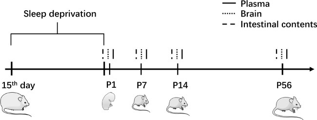
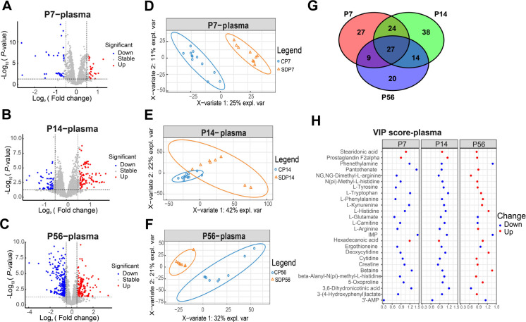
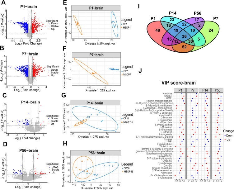
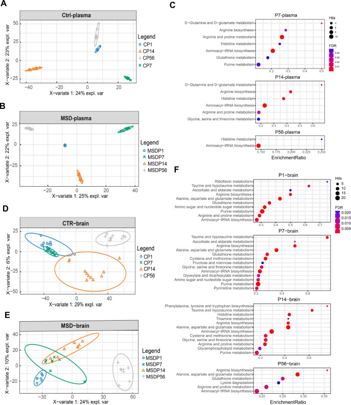
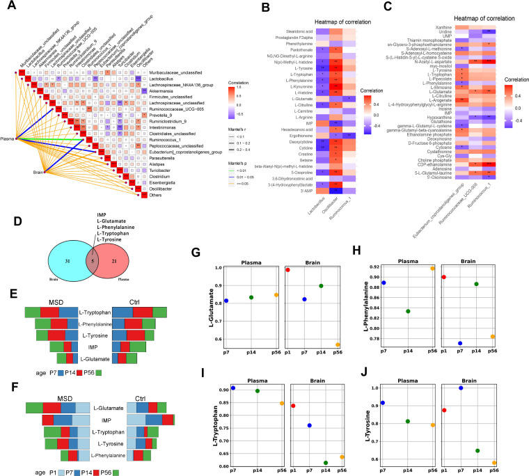
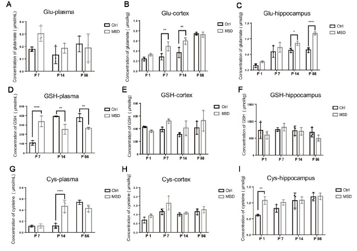
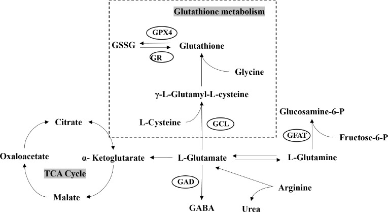
Maternal sleep deprivation (MSD) has emerged as a significant public health concern, yet its effects on offspring metabolism remain poorly understood. This study investigated the metabolomic implications of MSD on offspring cognitive development, with a particular focus on alterations in glutamate metabolism. Pregnant rats were subjected to sleep deprivation during late gestation. Plasma and brain samples from their offspring were collected at different postnatal days (P1, P7, P14, and P56) and analyzed using untargeted metabolomics with liquid chromatography-mass spectrometry. Metabolomic analysis revealed significant differences in various amino acids, including L-glutamate, L-phenylalanine, L-tyrosine, and L-tryptophan, which are crucial for cognitive function. Subsequent differential analysis and partial least squares discriminant analysis (sPLS-DA) demonstrated a gradual reduction in these metabolic differences in the brain as the offspring underwent growth and development. KEGG pathway analysis revealed differential regulation of several pathways, including alanine, aspartate, and glutamate metabolism, glutathione metabolism, arginine biosynthesis, aminoacyl-tRNA biosynthesis, histidine metabolism, and taurine and hypotaurine metabolism, at different developmental stages. Mantel and Spearman analyses indicated that the observed changes in metabolites in MSD progeny may be related to various gut microbes, Ruminococcus_1, Ruminococcaceae_UCG-005, and Eubacterium_coprostanoligenes_group. Biochemical assays further demonstrated developmental changes in the L-glutamate metabolic pathway. Collectively, these findings suggest that MSD not only affects maternal well-being but also has enduring metabolic consequences for offspring, particularly impacting pathways linked to cognitive function. This highlights the importance of addressing maternal sleep health to mitigate potential long-term consequences for offspring.

母体睡眠剥夺(MSD)已成为一个重要的社会问题,但其对后代代谢的影响仍知之甚少。该研究探讨了MSD对后代认知发育的代谢组学影响,特别是对谷氨酸代谢的改变。实验中,对妊娠晚期的大鼠进行睡眠剥夺处理,并在不同生后日(P1、P7、P14和P56)采集其子代的血浆和脑组织样本,通过液相色谱-质谱联用的非靶向代谢组学分析进行研究。代谢组学分析显示,L-谷氨酸、L-苯丙氨酸、L-酪氨酸和L-色氨酸等多种氨基酸存在显著差异,这些氨基酸对于认知功能至关重要。随后的差异分析和偏最小二乘判别分析(sPLS-DA)显示,随着子代的生长发育,这些代谢差异在脑组织中逐渐减少。KEGG通路分析揭示了不同发育阶段中,丙氨酸、天冬氨酸和谷氨酸代谢、谷胱甘肽代谢、精氨酸生物合成、氨酰tRNA生物合成、组氨酸代谢以及牛磺酸和亚牛磺酸代谢等多条通路的差异调控。Mantel检验和Spearman分析表明,MSD子代代谢物的变化可能与肠道微生物 Ruminococcus_1Ruminococcaceae_UCG-005Eubacterium_coprostanoligenes_group相关。生化分析进一步支持L-谷氨酸代谢途径的发育性变化。综上所述,这些发现表明,MSD不仅影响母体的健康,还对子代产生持久的代谢影响,尤其影响与认知功能相关的代谢途径。这凸显了关注母体睡眠健康的重要性,以减轻对后代潜在的长期影响。.

Keywords: Cognitive development; Glutamate metabolism; Maternal sleep deprivation; Metabolomics; Offspring.


Conflict of interest statement

The authors declare that they have no competing interests.

Figures

Figure 1
Figure 1
Animal model and sampling Pregnant rats were exposed to MSD for 6 h daily during gestation days 15–21, with prior acclimation to gentle handling procedures. Plasma, brain tissue, and intestinal content samples were collected from offspring on postnatal days 1, 7, 14, and 56 (P1, P7, P14, and P56, respectively).
Figure 2
Figure 2
Untargeted metabolomic analysis of plasma samples from offspring at different developmental stages A–C: Volcano plot of differential metabolites between MSD and control offspring at P7 (A), P14 (B), and P56 (C). D–F: sPLS-DA of metabolomes between MSD and control offspring at P7 (D), P14 (E), and P56 (F). G: Venn diagram of overlapping differential metabolites between MSD and control offspring at different time points in plasma. H: VIP scores of 27 common differential metabolites identified in the Venn diagram.
Figure 3
Figure 3
Untargeted metabolomic analysis of brain samples from offspring at different developmental stages A–D: Volcano plot of differential metabolites between MSD and control offspring at P1 (A), P7 (B), P14 (C), and P56 (D). E–H: sPLS-DA of metabolomes between MSD and control offspring at P1 (E), P7 (F), P14 (G), and P56 (H). I: Venn diagram of differential metabolites between MSD and control groups at different time points in the brain. J: VIP scores of 36 common differential metabolites identified in the Venn diagram.
Figure 4
Figure 4
sPLS-DA and pathway analysis of MSD and control offspring A–B: sPLS-DA plots of metabolomes in control (A) and MSD plasma (B) across all developmental stages. C: Graphical representation of pathway analysis of plasma samples (FDR<0.05). D, E: sPLS-DA plots of metabolomes in control (D) and MSD brain (E) across all developmental stages. F: Graphical representation of pathway analysis of brain samples (FDR<0.05).
Figure 5
Figure 5
Analysis of relationship between metabolites and gut microbiota A: Mantel analysis of correlation between common differential metabolites in plasma and brain with gut microbiota. B, C: Spearman analysis of correlation between identified gut microbiota from Mantel test and common differential metabolites in plasma (B) and brain samples (C). *: P<0.05; **: P<0.001; ***: P<0.0001. D: Venn diagram showcasing common differential metabolites in both brain and plasma. E, F: Comparative analysis of concentrations of five common differential metabolites in plasma (E) and brain samples (F) between MSD and control groups. G–J: Dots, with colors corresponding to AUCs depicted in Supplementary Figures S3, S4, signify areas under the ROC curves for metabolites exhibiting biomarker potential for MSD-exposed offspring. Ctrl: Control group. MSD: Maternal sleep deprivation group.
Figure 6
Figure 6
Biochemical assessment of glutamate, glutathione, and cysteine levels A–C: Glutamate concentrations in plasma (A), cortex (B), and hippocampus (C). D–F: Glutathione concentrations in plasma (D), cortex (E), and hippocampus (F). G–I: Cysteine concentrations in plasma (G), cortex (H), and hippocampus (I). Glu: Glutamate; GSH: Glutathione; Cys: Cysteine. Ctrl: Control group. MSD: Maternal sleep deprivation group. *: P<0.05; **: P<0.001.
Figure 7
Figure 7
Overview of glutamate and glutathione metabolism GSSG: Glutathione disulfide; GABA: Gamma-aminobutyric acid; GPX4: Glutathione peroxidase 4; GR: Glutathione reductase; GCL: γ-glutamylcysteine ligase; GFAT: Glutamine fructose-6-phosphate amidotransferase; GAD: Glutamate decarboxylase; TCA: Tricarboxylic acid cycle.

References

    1. Alvarenga TA, Aguiar MFP, Mazaro-Costa R, et al Effects of sleep deprivation during pregnancy on the reproductive capability of the offspring. Fertility and Sterility. 2013;100(6):1752–1757. doi: 10.1016/j.fertnstert.2013.08.014. - DOI - PubMed
    1. Aswathy BS, Kumar VM, Gulia KK The effects of rapid eye movement sleep deprivation during late pregnancy on newborns' sleep. Journal of Sleep Research. 2018;27(2):197–205. doi: 10.1111/jsr.12564. - DOI - PubMed
    1. Banks S, Dinges DF Behavioral and physiological consequences of sleep restriction. Journal of Clinical Sleep Medicine. 2007;3(5):519–528. doi: 10.5664/jcsm.26918. - DOI - PMC - PubMed
    1. Biagi E, Franceschi C, Rampelli S, et al Gut microbiota and extreme longevity. Current Biology. 2016;26(11):1480–1485. doi: 10.1016/j.cub.2016.04.016. - DOI - PubMed
    1. Chakraborty P, Dey A, Gopalakrishnan AV, et al Glutamatergic neurotransmission: a potential pharmacotherapeutic target for the treatment of cognitive disorders. Ageing Research Reviews. 2023;85:101838. doi: 10.1016/j.arr.2022.101838. - DOI - PubMed