. 2024 Nov 13;15(11):e0232724.
doi: 10.1128/mbio.02327-24. Epub 2024 Oct 9.

Human parainfluenza virus 3 field strains undergo extracellular fusion protein cleavage to activate entry

Affiliations

Human parainfluenza virus 3 field strains undergo extracellular fusion protein cleavage to activate entry

Kyle Stearns et al. mBio. .

Abstract

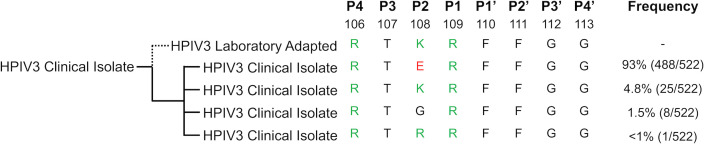
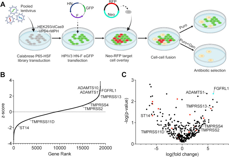
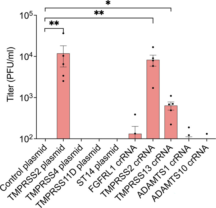
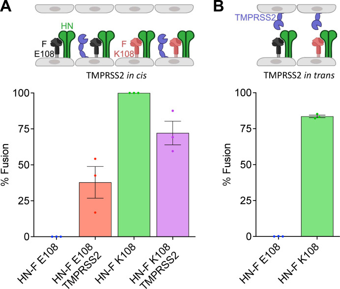
Human parainfluenza virus 3 (HPIV3) infection is driven by the coordinated action of viral surface glycoproteins hemagglutinin-neuraminidase (HN) and fusion protein (F). Receptor-engaged HN activates F to insert into the target cell membrane and drive virion-cell membrane fusion. For F to mediate entry, its precursor (F0) must first be cleaved by host proteases. F0 cleavage has been thought to be executed during viral glycoprotein transit through the trans-Golgi network by the ubiquitously expressed furin because F0 proteins of laboratory-adapted viruses contain a furin recognition dibasic cleavage motif RXKR around residue 108. Here, we show that the F proteins of field strains have a different cleavage motif from laboratory-adapted strains and are cleaved by unidentified proteases expressed in only a narrow subset of cell types. We demonstrate that extracellular serine protease inhibitors block HPIV3 F0 cleavage for field strains, suggesting F0 cleavage occurs at the cell surface facilitated by transmembrane proteases. Candidate proteases that may process HPIV3 F in vivo were identified by a genome-wide CRISPRa screen in HEK293/dCas9-VP64 + MPH cells. The lung-expressed extracellular serine proteases TMPRSS2 and TMPRSS13 are both sufficient to cleave HPIV3 F and enable infectious virus release by otherwise non-permissive cells. Our findings support an alternative mechanism of F activation in vivo, reliant on extracellular membrane-bound serine proteases expressed in a narrow subset of cells. The proportion of HPIV3 F proteins cleaved and infectious virus release is determined by host cell expression of requisite proteases, allowing just-in-time activation of F and positioning F cleavage as another key regulator of HPIV3 spread.

Importance: Enveloped viruses cause a wide range of diseases in humans. At the first step of infection, these viruses must fuse their envelope with a cell membrane to initiate infection. This fusion is mediated by viral proteins that require a critical activating cleavage event. It was previously thought that for parainfluenza virus 3, an important cause of respiratory disease and a representative of a group of important pathogens, this cleavage event was mediated by furin in the cell secretory pathways prior to formation of the virions. We show that this is only true for laboratory strain viruses, and that clinical viruses that infect humans utilize extracellular proteases that are only made by a small subset of cells. These results highlight the importance of studying authentic clinical viruses that infect human tissues for understanding natural infection.

Keywords: fusion protein; membrane fusion; parainfluenza virus; paramyxovirus; proteases; viral entry.


Conflict of interest statement

K.S., M.P., and A.M. are listed as inventors on a provisional patent related to the work in this article.

Figures

Fig 1
Fig 1
Human parainfluenza viruses in circulation have different fusion protein cleavage motifs than lab-adapted strains. HPIV3 fusion protein sequences were extracted with NCBI Protein Blast [NC_001796 fusion protein (NP_067151.1, accessed 5/18/22)]. Listed are cleavage motif sequence alignments of laboratory-adapted and patient-derived HPIV3. Reported are the observed sequence frequencies and the amino acid sequences of four amino acids upstream and downstream of the HPIV3 F1/F2 cleavage site. Green, positively charged amino acids; red, negatively charged amino acids.
Fig 2
Fig 2
HPIV3 fusion protein cleavage and infectious virion production by ex vivo tissues and immortalized cell cultures. The indicated tissue (HAE) and immortalized cells were inoculated with HPIV3 F E108 (left) or HPIV3 F K108 (right) with the JS strain background. HPIV3 in cell culture media and HAE apical washes were collected 1–3 days after infection. (A) HPIV3-infected cell culture media and HAE apical wash resolved by reducing SDS-PAGE and immunoblotting with anti-HPIV3 F antibody. (B) HPIV3 titer in cell culture media and HAE apical washes 1–3 days after infection. (C) Proteases with transcripts per million (TPM) expression level >5 in HAE or Calu-3, and <5 in A549, HEK293T, and HepG2 cells determined by RNA-seq. Values are means and standard error of the means from three biological replicates.
Fig 3
Fig 3
Inhibition of extracellular serine proteases with aprotinin or leupeptin blocks HPIV3 F (E108) cleavage and reduces the production of infectious HPIV3. Calu-3 cells were inoculated with HPIV3 F E108 (red) or HPIV3 F K108 (blue) with JS strain background. After inoculation, cells were treated with the indicated concentrations of aprotinin or leupeptin. (A) HPIV3-infected Calu-3 cell culture media were resolved by reducing SDS-PAGE and immunoblotted with anti-HPIV3 F antibody. (B) HPIV3 titer in cell culture media was assessed 3 days after infection. Values are means and SEM from three biological replicates.
Fig 4
Fig 4
Inhibition of extracellular HPIV3 F (E108) cleavage by aprotinin or steric hindrance of the F cleavage site on HAE. HAE cells were inoculated with HPIV3 F E108 or HPIV3 F K108 with CI-1 background. After inoculation, cells were apically treated with vehicle, 1 mM aprotinin, or 2.6 µM 4C06 VHH-Fc. (A) HPIV3-infected HAE supernatants were resolved by reducing SDS-PAGE and immunoblotted with anti-HPIV3 F antibody. (B) Proportion of HPIV3 F cleaved. ****P ≤ 0.0001 by two-way analysis of variance (ANOVA) and Tukey’s multiple comparisons post hoc test. (C) HAE supernatant HPIV3 titered in Vero cells infected in the presence or absence of 1 µg/mL TPCK-treated trypsin. **P ≤ 0.01 by two-way ANOVA and Sidak’s post hoc test. Values are means and SEM from three biological replicates.
Fig 5
Fig 5
Whole human genome CRISPRa screen identified candidate proteases for HPIV3 F cleavage and infection. (A) Schematic of whole human genome lentiviral screen (created with BioRender.com). (B) Distribution of normalized z-scores (geneticin treated vs control) for all genes targeted by the sgRNA library. (C) Volcano plot of all human proteases expressed in human lung airway epithelium with serine proteases indicated in red. FGFRL1 was included as a positive control (turquoise).
Fig 6
Fig 6
CRISPRa gain-of-function screen identifies TMPRSS2 and TMPRSS13 as sufficient for HPIV3 infection. HEK293/dCas9-VP64 + MPH cells were transfected with CRISPRa plasmids or tracrRNA:crRNA complexes for proteases significantly enriched in the lentiviral screen. Transfected cells were then infected with HPIV3 F E108 with JS strain background and infectious HPIV3 released by cells were titered. *P ≤ 0.05, **P ≤ 0.01 by two-way analysis of variance and Kruskal-Wallis post hoc test. Values are means and SEM from three biological replicates.
Fig 7
Fig 7
TMPRSS2 expression is sufficient for HPIV3 HN-F-mediated cell-cell fusion. Fusion activity measured with β-galactosidase complementation assay. HEK293T cells were transfected with the α subunit of β-galactosidase and HPIV3 F E108/HN (blue), HPIV3 F E108/HN/TMPRSS2 (red), HPIV3 F K108/HN (green), or HPIV3 F K108/HN/TMPRSS2 (purple). (A) Cells were incubated for 16 hours with HEK293T cells expressing the Ω subunit of β-galactosidase. (B) Cells were then incubated for 16 hours with HEK293T-TMPRSS2 (TMPRSS2 in trans) cells expressing the Ω subunit of β-galactosidase. Values are means and SEM from three biological replicates. Schematic created with BioRender.com.
Fig 8
Fig 8
TMPRSS2 and TMPRSS13 are not necessary for infectious viral particle production by Calu-3 cells. WT, TMPRSS2 KO, TMPRSS13 KO, and TMPRSS2/13 KO Calu-3 cells were inoculated with HPIV3 F E108 or HPIV3 F K108 with JS strain background. HPIV3 titers in Calu-3 culture media. Values are means and SEM from three to six biological replicates.

References

    1. Nair H, Nokes DJ, Gessner BD, Dherani M, Madhi SA, Singleton RJ, O’Brien KL, Roca A, Wright PF, Bruce N, Chandran A, Theodoratou E, Sutanto A, Sedyaningsih ER, Ngama M, Munywoki PK, Kartasasmita C, Simões EAF, Rudan I, Weber MW, Campbell H. 2010. Global burden of acute lower respiratory infections due to respiratory syncytial virus in young children: a systematic review and meta-analysis. Lancet 375:1545–1555. doi:10.1016/S0140-6736(10)60206-1 - DOI - PMC - PubMed
    1. Williams JV, Harris PA, Tollefson SJ, Halburnt-Rush LL, Pingsterhaus JM, Edwards KM, Wright PF, Crowe JE. 2004. Human metapneumovirus and lower respiratory tract disease in otherwise healthy infants and children. N Engl J Med 350:443–450. doi:10.1056/NEJMoa025472 - DOI - PMC - PubMed
    1. Weinberg GA, Hall CB, Iwane MK, Poehling KA, Edwards KM, Griffin MR, Staat MA, Curns AT, Erdman DD, Szilagyi PG, New Vaccine Surveillance Network . 2009. Parainfluenza virus infection of young children: estimates of the population-based burden of hospitalization. J Pediatr 154:694–699. doi:10.1016/j.jpeds.2008.11.034 - DOI - PubMed
    1. Black RE, Cousens S, Johnson HL, Lawn JE, Rudan I, Bassani DG, Jha P, Campbell H, Walker CF, Cibulskis R, Eisele T, Liu L, Mathers C. 2010. Global, regional, and national causes of child mortality in 2008: a systematic analysis. The Lancet 375:1969–1987. doi:10.1016/S0140-6736(10)60549-1 - DOI - PubMed
    1. Moscona A. 2005. Entry of parainfluenza virus into cells as a target for interrupting childhood respiratory disease. J Clin Invest 115:1688–1698. doi:10.1172/JCI25669 - DOI - PMC - PubMed

LinkOut - more resources