. 2024 Dec;103(12):104238.
doi: 10.1016/j.psj.2024.104238. Epub 2024 Sep 2.

Up-regulated Lnc BTU promotes the production of duck plague virus DNA polymerase and inhibits the activation of JAK-STAT pathway to facilitate duck plague virus replication

Affiliations

Up-regulated Lnc BTU promotes the production of duck plague virus DNA polymerase and inhibits the activation of JAK-STAT pathway to facilitate duck plague virus replication

Ning Luo et al. Poult Sci. 2024 Dec.

Abstract

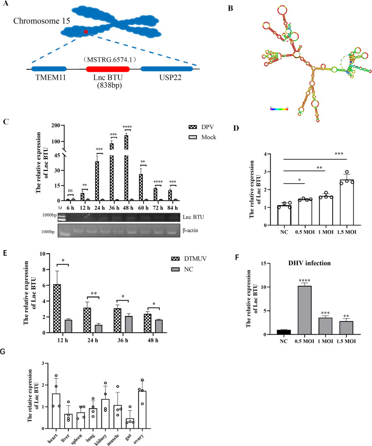
Duck plague virus (DPV) is the only herpes virus known to be transmissible among aquatic animals, leading to immunosuppression in ducks, geese and swans. Long noncoding RNAs (LncRNA) are known to participate in viral infections, acting as either immune defenders or viral targets to evade the host response, but their precise roles in waterfowl virus infections are yet to be fully understood. This study aimed to investigate the role of LncRNA in DPV-induced innate immune responses. Results showed that DPV infection greatly upregulated Lnc BTU expression in duck embryo fibroblasts (DEF) and Lnc BTU promoted DPV replication. Mechanically, 4 DPV proteins, namely UL46, UL42, VP22 and US10, interacted with Lnc BTU, leading to its upregulation. Specifically, Lnc BTU facilitated the production of DNA polymerase by enhancing UL42 expression, thereby promoting DPV replication. Additionally, Lnc BTU suppressed STAT1 expression by targeting the DNA binding domain (DBD) and promoting STAT1 degradation through the proteasome pathway. Furthermore, Lnc BTU inhibited the production of key antiviral factors such as IFN-α, IFN-β, MX and OASL during DPV infection. Treatment with 2 JAK-STAT pathway activators in DEFs resulted in the inhibition of Lnc BTU expression and DPV replication. Interestingly, DPV infection led to a decrease in STAT1 levels, which was reversed by Si-Lnc BTU. These findings suggest that DPV relies on Lnc BTU to inhibit the activation of the JAK-STAT pathway and limit the production of type 1 interferons (IFN) to complete immune evasion. Our study highlights the novel role of DPV proteins UL46, UL42, VP22, US10 as RNA-binding proteins in modulating the innate antiviral immune response, and discover the role of a new host factor, Lnc BTU, in DPV immune evasion, Lnc BTU and STAT1 can be used as a potential therapeutic target for DPV infection and immune evasion.

Keywords: Duck plague virus; JAK-STAT pathway; Lnc BTU; STAT1; innate immunity.


Conflict of interest statement

DISCLOSURES The authors declare that they have no known competing financial interests or personal relationships that could have appeared to influence the work reported in this paper.

Figures

Figure 1
Figure 1
Bioinformatics analysis and expression kinetics analysis of Lnc BTU. (A) Schematic diagram of the location of Lnc BTU on chromosomes in the duck genome. (B) RNA secondary structure prediction for Lnc BTU was analyzed using RNA-fold (http://rna.tbi.univie.ac.at/cgi-bin/RNAWebSuite/RNAfold.cgi) and the data were shown as a minimal free energy structure (MFE = −240.25 kcal/mol). Base pairing probabilities have been color-coded from 0 (blue) to 1 (red). (C) Lnc BTU mRNA expression detected by qRT-PCR and PCR in DEFs infected DPV (MOI = 1) at indicate time. (D) Lnc BTU mRNA expression detected by qRT-PCR in DEFs infected DPV (MOI = 0.5, 1, 1.5). (E, F) Lnc BTU mRNA expression detected by qRT-PCR in DEFs infected DTMUV or DHV. (G) QRT-PCR detected Lnc BTU in normal duck tissues. (Each experiment was repeated 3 times. ns, P>0.05; *, P < 0.05; **, P < 0.01; ***, P < 0.001; ****, P < 0.0001).
Figure 2
Figure 2
Lnc BTU promotes DPV infection in DEFs. (A, B) QRT-PCR and PCR confirmed overexpression or knockdown Lnc BTU efficiency. (C-D) pCAGGS-Lnc BTU (pCAGGS as control) or Si-1 (Si-NC as control) transfected DEFs after DPV infection and DPV copy number in the supernatant was measured using qRT-PCR at indicate time. (E) DPV titer was detected using TCID50 assay. (Each experiment was repeated 3 times. ns, P > 0.05; *, P < 0.05; **, P < 0.01; ***, P < 0.001; ****, P < 0.0001.)
Figure 3
Figure 3
Lnc BTU directly interacts with DPV UL46, UL42, VP22 and US10. (A) Coomassie brilliant blue assay detect Lnc BTU enriched protein. DEFs were transfected with pCAGGS-F2-LncBTU or pCAGGS-F2 (control) for 24 h, then infected with DPV at MOI = 1 for 24 h, the cells were harvested and followed by Western blot. (B, C, D) MS Analysis, host proteins in pCAGGS-F2-lnc BTU group or pCAGGS-F2-Lnc BTU+DPV group obtained by STRING software. (E–H) RNA pull-down assay was detected Lnc BTU interact with viral protein. HEK293T cells were transfected with pCAGGS-F2-LncBTU and viral protein plasmid (pCAGGS-UL46-flag, pCAGGS-UL42-Flag plasmid, pCAGGS-VP22-HA plasmid and pCAGGS-US10-Flag plasmid respectively) for 36 h, followed by Western blot with RNA pull-down kit manual. (I–L) RIP assay to detected 4 viral proteins interact with Lnc BTU. DEFs were transfected 4 viral plasmids respectively and pCAGGS-Lnc BTU for 36 h, the cells were harvested and Lnc BTU mRNA level detected by PCR and qRT-PCR. Each experiment was repeated 3 times. ns, P > 0.05; *, P < 0.05; **, P < 0.01, ***, P < 0.001; ****, P < 0.0001.
Figure 4
Figure 4
Lnc BTU promoted UL46, UL42 and VP22 expression, inhibited US10 expression and UL46, UL42, VP22 and US10 all promoted Lnc BTU expression. (A–D) DEFs were transfected with pCAGGS-Lnc BTU for 36 h, then the cell infected with DPV at MOI = 1. QRT-PCR measured UL46, UL42, VP22 and US10 mRNA level at indicate time. (E–H) doses of pCAGGS-Lnc BTU DEFs were transfected with pCAGGS-UL46-Flag (E), pCAGGS-UL42-Flag (F), pCAGGS-VP22-HA (G) or pCAGGS-US10-Flag (H) for 36h, the cell lysates were subjected to Western blotting. (I–L) Doses Si-Lnc BTU DEFs were transfected with pCAGGS-UL46-Flag (I) or pCAGGS-UL42-Flag (J) or pCAGGS-VP22-HA (K) or pCAGGS-US10-Flag (L) for 36h, the cell lysates were subjected to Western blotting. The resulting band plot was examined for gray value by using image-J software. Bar graphs represent the mean of 3 independent experiments (±SD). (M) To explore UL46, UL42, VP22 and US10 effect on Lnc BTU expression, doses of pCAGGS-UL46-Flag, pCAGGS-UL42-Flag, pCAGGS-VP22-HA, pCAGGS-US10-Flag transfected separately to DEFs for 36 h, qRT-PCR to detect Lnc BTU mRNA level. (N) pCAGGS-Lnc BTU DEFs transfected doses of pCAGGS-UL46-Flag, pCAGGS-UL42-Flag, pCAGGS-VP22-HA or pCAGGS-US10-Flag and transfected to DEFs for 36 h, qRT-PCR to detect Lnc BTU mRNA level. Each experiment was repeated 3 times. ns, P > 0.05; *, P < 0.05; **, P < 0.01, ***, P < 0.001; ****, P < 0.0001.
Figure 4
Figure 4
Lnc BTU promoted UL46, UL42 and VP22 expression, inhibited US10 expression and UL46, UL42, VP22 and US10 all promoted Lnc BTU expression. (A–D) DEFs were transfected with pCAGGS-Lnc BTU for 36 h, then the cell infected with DPV at MOI = 1. QRT-PCR measured UL46, UL42, VP22 and US10 mRNA level at indicate time. (E–H) doses of pCAGGS-Lnc BTU DEFs were transfected with pCAGGS-UL46-Flag (E), pCAGGS-UL42-Flag (F), pCAGGS-VP22-HA (G) or pCAGGS-US10-Flag (H) for 36h, the cell lysates were subjected to Western blotting. (I–L) Doses Si-Lnc BTU DEFs were transfected with pCAGGS-UL46-Flag (I) or pCAGGS-UL42-Flag (J) or pCAGGS-VP22-HA (K) or pCAGGS-US10-Flag (L) for 36h, the cell lysates were subjected to Western blotting. The resulting band plot was examined for gray value by using image-J software. Bar graphs represent the mean of 3 independent experiments (±SD). (M) To explore UL46, UL42, VP22 and US10 effect on Lnc BTU expression, doses of pCAGGS-UL46-Flag, pCAGGS-UL42-Flag, pCAGGS-VP22-HA, pCAGGS-US10-Flag transfected separately to DEFs for 36 h, qRT-PCR to detect Lnc BTU mRNA level. (N) pCAGGS-Lnc BTU DEFs transfected doses of pCAGGS-UL46-Flag, pCAGGS-UL42-Flag, pCAGGS-VP22-HA or pCAGGS-US10-Flag and transfected to DEFs for 36 h, qRT-PCR to detect Lnc BTU mRNA level. Each experiment was repeated 3 times. ns, P > 0.05; *, P < 0.05; **, P < 0.01, ***, P < 0.001; ****, P < 0.0001.
Figure 5
Figure 5
Lnc BTU enhances the synthesis of DPV DNA polymerase. (A) DEFs infected DPV at MOI=1, then treated with Cidofovir (10 nmol) for 1 h, 2 h, 3 h, 4 h, 6 h, 8 h, 10 h, 12 h, 14 h and 16 h, qRT-PCR detected DPV copy numbers in the supernatant. (B, C) DEFs were transfected pCAGGS-UL30-HA and doses of pCAGGS-Lnc BTU or Si-Lnc BTU for 36 h. cell harvested for Western blotting, and the resulting band plot gray value was examined using image-J software. (D, E) DEFs co-transfected pCAGGS-UL42-Flag, pCAGGS-UL30-HA and doses of pCAGGS-Lnc BTU or Si-Lnc BTU for 36 h, Co-IP detected UL30 and UL42 protein expression.
Figure 6
Figure 6
Lnc BTU inhibits the activation of the JAK-STAT signaling pathway by targeting STAT1 (A) Doses Poly (I:C) treated DEFs for 36 h, qRT-PCR and PCR detected Lnc BTU expression. (B) pCAGGS-Lnc BTU treated DEFs for 24 h, then infected DPV at MOI = 1.5 for 12 h, 24 h, 36 h and 48 h, qRT-PCR detected IFN-β level. (C) HEK293T cells were co-transfected with pCAGGS-STAT1-Myc and pCAGGS-F2-LncBTU for 36 h, cell harvested and followed by RNA pull-down kit manual. (D) RIP assay to detected STAT1 interact with Lnc BTU. DEFs co-transfected with pCAGGS-F2-LncBTU and pCAGGS-STAT1-Myc for 36 h, the cells were harvested and Lnc BTU mRNA level detected by PCR and qRT-PCR. (E, F) DEFs transfected with doses of Si-Lnc BTU (E) (0 nmol, 10 nmol, 20 nmol, 30 nmol, 50 nmol, 100 nmol) or pCAGGS-Lnc BTU (F) (0 μg, 0.5 μg, 1 μg, 1.5 μg, 2 μg, 3 μg) for 24h, infected 1.5 MOI DPV, qRT-PCR detected STAT1 mRNA level. (G, H) doses of Si-Lnc BTU (G) or pCAGGS-Lnc BTU (H) DEFs transfected with pCAGGS-STAT1-Myc for 36h, STAT1 was detected by Western blotting. (I–T) IFN-α, IFN-β, MX, OASL, IL-6, IRF7 mRNA level in doses of Si-Lnc BTU (0nmol, 10nmol, 20nmol, 30nmol, 50nmol, 100nmol) or pCAGGS-Lnc BTU (0μg, 0.5μg, 1μg, 1.5μg, 2μg, 3μg) DEFs after DPV infection (MOI = 1.5) measured by qRT-PCR, using β-actin as internal control. Each experiment was repeated 3 times. ns, P > 0.05; *, P < 0.05; **, P < 0.01; ***, P < 0.001; ****, P < 0.0001.
Figure 7
Figure 7
(A) QRT-PCR was used to detect STAT1 mRNA level at 6 h, 12 h, 24 h, 36 h, 48 h and 60 h after 1.5MOI DPV infection. (B) Si-Lnc BTU transfected to DEFs for 24 h, then infected DPV at MOI = 1.5 for 24 h, qRT-PCR detected STAT1 level. (C, D) DEFs were pretreated with RO8191 (5 μmol/mL), 2-NP (5 μmol/mL) or solvent DMSO for 1 h, and then infected with 1.5 MOI DPV for another 1 h, JAK1, JAK2, TYK2, STAT1, STAT2, IFN-α, IFN-β, IRF7, MX, OASL and IL-6 levels and DPV copy numbers were detected by qRT-PCR. (E) RO8191 (5 μmol/mL) and 2-NP (5 μmol/mL) treated DEFs for 2 h, then infected DPV at MOI = 1.5 for 6 h, qRT-PCR detected Lnc BTU level. (F) pCAGGS-STAT1 DEFs infected DPV at MOI = 1, qRT-PCR detected DPV copy numbers at indicate time. (G–J) DEFs transfected with pCAGGS-STAT1 or si-STAT1 for 24 h, then infected DPV at MOI = 1, qRT-PCR detected UL46, UL42, VP22 and US10 level at indicate time.
Figure 8
Figure 8
Lnc BTU inhibits STAT1 expression in the nucleus and medicates STAT1 degradation via the ubiquitin-proteasome pathway. (A) STAT1 domains and truncation mutants. (B–D) HEK293T cells co-transfected with pCAGGS-F2-LncBTU and pCAGGS-STAT1-△SH2-Myc (B) or pCAGGS-STAT1-△DBD-Myc (C) or pCAGGS-STAT1-CCD-Myc (D) for 36 h, cells harvested and followed by RNA pull-down kit manual and Western blotting. (E–G) RIP assay to detected STAT1 truncation mutants interact with Lnc BTU. DEFs transfected with pCAGGS-F2-LncBTU and pCAGGS-STAT1-△SH2-Myc (E), pCAGGS-STAT1-△DBD-Myc (F) or pCAGGS-STAT1-CCD-Myc (G) for 36 h, cells harvested and used to PCR and qRT-PCR. (H) DEFs were infected with 1.5 MOI DPV for 36 h, qRT-PCR detected Lnc BTU expression obtained following nucleoplasmic isolation manual. (I, J) Si-Lnc BTU or pCAGGS-Lnc BTU DEFs infected 1.5 MOI DPV for 24 h, qRT-PCR (I) and Western blotting (J) detected STAT1 expression following nucleoplasmic isolation manual. (K, L) The colocalization between the Lnc BTU and STAT1 in DPV-infected DEFs detected by FISH assays. (M) DEFs, transfected with pCAGGS-Lnc BTU and pCAGGS-STAT1-Myc for 36 h, were treated with CHX (10 nmol/ml) at indicated times, followed by Western blotting. (N) DEFs transfected with pCAGGS-STAT1-Myc and pCAGGS-Lnc BTU for 36 h, then treated with MG132 (5 nmol/ml) or Bafilomycin A1 (10 nmol/ml) or CQ (10 nmol/ml) for 6 h, STAT1 protein expression detected by Western blotting. The resulting band plot gray value was examined by using image-J software.
Figure 9
Figure 9
Schematic diagram of Lnc BTU targeting STAT1 in the JAK-STAT signaling pathway to help DPV evade host innate immunity.

Similar articles

Cited by

References

    1. Apinda N., Muenthaisong A., Chomjit P., Sangkakam K., Nambooppha B., Rittipornlertrak A., Koonyosying P., Yao Y., Nair V., Sthitmatee N. Simultaneous protective immune responses of Ducks against Duck plague and fowl cholera by recombinant duck enteritis virus vector expressing pasteurella multocida OmpH gene. Vaccines. 2022;10:1358. - PMC - PubMed
    1. Apinda N., Yao Y., Zhang Y., Reddy V.R.A.P., Chang P., Nair V., Sthitmatee N. CRISPR/Cas9 editing of Duck enteritis virus genome for the construction of a recombinant vaccine vector expressing omph gene of pasteurella multocida in two novel insertion sites. Vaccines. 2022;10:686. - PMC - PubMed
    1. Burrel S., Aït-Arkoub Z., Agut H., Boutolleau D. Genotypic characterization of herpes simplex virus DNA polymerase UL42 processivity factor. Antiviral. Res. 2012;93:199–203. - PubMed
    1. Cai B., Cai J., Luo Y., Chen C., Zhang S. The Specific roles of JAK/STAT signaling pathway in sepsis. Inflammation. 2015;38:1599–1608. - PubMed
    1. Cai R., Sun Y., Qimuge N., Wang G., Wang Y., Chu G., Yu T., Yang G., Pang W. Adiponectin AS lncRNA inhibits adipogenesis by transferring from nucleus to cytoplasm and attenuating adiponectin mRNA translation. Biochim. Biophys. Acta. Mol. Cell. Biol. Lipids. 2018;1863:420–432. - PubMed

MeSH terms

Supplementary concepts