. 2024 Nov;300(11):107870.
doi: 10.1016/j.jbc.2024.107870. Epub 2024 Oct 9.

Redox control of the deubiquitinating enzyme Ubp2 regulates translation during stress

Affiliations

Redox control of the deubiquitinating enzyme Ubp2 regulates translation during stress

Clara M Santos et al. J Biol Chem. 2024 Nov.

Abstract

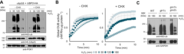
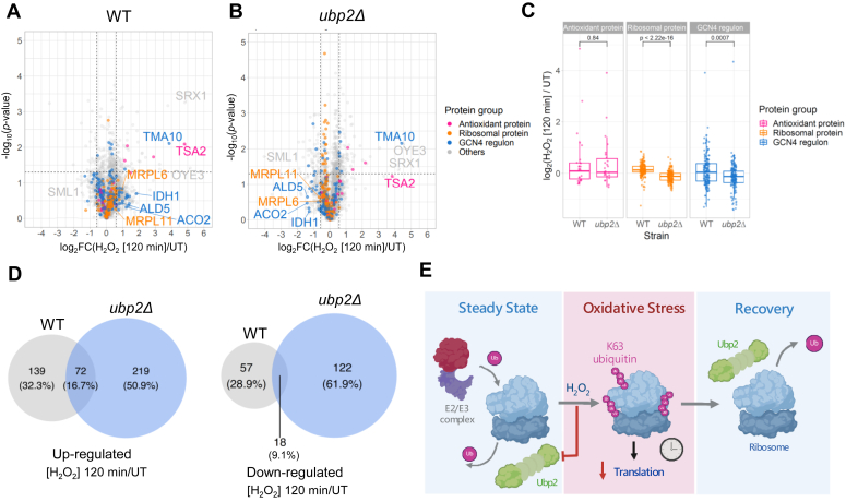
Protein ubiquitination is essential to govern cells' ability to cope with harmful environments by regulating many aspects of protein dynamics from synthesis to degradation. As important as the ubiquitination process, the reversal of ubiquitin chains mediated by deubiquitinating enzymes (DUBs) is critical for proper recovery from stress and re-establishment of proteostasis. Although it is known that ribosomes are decorated with K63-linked polyubiquitin chains that control protein synthesis under stress, the mechanisms by which these ubiquitin chains are reversed and regulate proteostasis during stress recovery remain elusive. Here, we showed in budding yeast that the DUB Ubp2 is redox-regulated during oxidative stress in a reversible manner, which determines the levels of K63-linked polyubiquitin chains present on ribosomes. We also demonstrate that Ubp2 can cleave single ubiquitin moieties out of chains and its activity is modulated by a series of repeated domains and the formation of disulfide bonds. By combining cellular, biochemical, and proteomics analyses, we showed that Ubp2 is crucial for restoring translation after stress cessation, indicating an important role in determining the cellular response to oxidative stress. Our work demonstrates a novel role for Ubp2, revealing that a range of signaling pathways can be controlled by redox regulation of DUB activity in eukaryotes, which in turn will define cellular states of health and diseases.

Keywords: deubiquitylation (deubiquitination); oxidative stress; redox regulation; translation control; yeast.


Conflict of interest statement

Conflict of interest The authors declare that they have no conflicts of interest with the contents of this article.

Figures

Figure 1
Figure 1
Ubp2 promotes K63-ub cleavage after stress and is reversibly inhibited by H2O2.A, deletion and mutation of UBP2 impairs K63-ub chain removal from ribosomes. Immunoblot anti-K63-ub of isolated ribosomes from cells treated in the presence or absence of 0.6 mM of H2O2 for 30 min (H2O2), followed by 20 min recovery (Rec) in fresh medium. Anti-uL5/Rpl11 was used as loading control for isolated ribosomes and anti-HA for Ubp2 (n = 2). B, Ubp2 preferentially cleaves K63-ub chains. Immunoblot anti-ub of synthetic tetra K63-ub (250 ng) or K48-ub chains (250 ng) incubated in the presence or absence of 3 μg of purified Ubp2 or pan DUB USP2 (n = 2). C, immunoblot anti-ub of synthetic K63-ub chains (250 ng) incubated in the presence or absence of 3 μg of purified Ubp2. Arrows indicate degradation products (n = 2). D, Ubp2’s activity is redox-regulated in vitro. Activity of recombinant Ubp2 (70 ng) was assessed in vitro using 0.75 μM of Ub-Rho fluorophore. Ubp2 was incubated for 5 min with 100 μM or 500 μM of H2O2, 20 mM IAA (cysteine alkylator), or 100 μM PR-619 (general DUB inhibitor). Two millimolars of DTT (reducing agent) was added after 5 min of assay to restore Ubp2 activity. Rho 110 fluorescence was recorded at 535 nm with excitation at 485 nm (n = 2, two technical replicates each). E, Ubp2 deubiquitinating activity is impaired by H2O2. Immunoblot anti-ub of synthetic tetra K63-ub chains (250 ng) incubated in the presence or absence of 3 μg purified Ubp2 with the addition or absence of 7 mM H2O2, 10 mM DTT, or 10 mM IAA (n = 3). F, Ubp2 activity is redox-regulated in yeast cells under stress. Ubp2-HA and Ubp2C745S-HA were immunoprecipitated from yeast cells untreated or treated with 0.6 mM H2O2 for 30 min. Ubp2 activity was assessed in vitro using 1.5 μM of Ub-AMC fluorophore. 20 mM of DTT was added after 60 min as indicated by the arrow to restore Ubp2 activity. Fluorescence was recorded at 445 nm with excitation at 345 nm (n = 2). Reactions shown in (B, C, and E) were incubated for 1 h at 30 °C prior SDS-PAGE and immunoblotting. DUB, deubiquitinating enzyme; H2O2, hydrogen peroxide; HA, hemagglutinin; IAA, iodoacetamide; K63-ub, K63-linked polyubiquitin; MW, molecular weight; Ub, ubiquitin.
Figure 2
Figure 2
Ubp2 is sensitive to H2O2.A, accumulation of K63-ub chains is differentially induced by H2O2 and organic peroxides. Immunoblot anti-K63-ub from WT cells exposed to 0.6 mM and 2.5 mM of H2O2, or organic peroxides tert-butyl (t-BHP) and cumene hydroperoxide (CHP) for 30 min. Anti-PGK1 was used as a loading control (n = 3). B, cellular deubiquitinating activity is differentially affected by H2O2 and organic peroxides. WT cells untreated (UT) or treated with either 0.6 mM (left) or 2.5 mM (right) of peroxides (ROOH) for 30 min were lysed, and total DUB activity was determined by incubating the whole-cell extract with the fluorogenic substrate Ub-Rho. Total DUB activity was assessed over time using 0.75 μM of Ub-Rho, whose fluorescence was determined at 535 nm with excitation at 485 nm. Signal was normalized by protein concentration and a standard curve of free rhodamine 110 (n = 2, two technical replicates each). C and D, Ubp2 and CezanneCAT are sensitive to H2O2. C, purified Ubp2 (70 ng) and D, CezanneCAT (3 μg) were incubated with 500 μM H2O2, t-BHP, and CHP for 5 min and DUB activity was assessed as described above. Activity was normalized by Rho 110 fluorescence registered for untreated Ubp2 or CezanneCAT. Bar graphs show mean values ± SD for three biological replicates. CezanneCAT activity was performed in duplicate and error bars represent standard error. Significance was calculated using an unpaired two-tailed Student’s t test, where ∗p < 0.05 and ∗∗p < 0.005. DUB, deubiquitinating enzyme; H2O2, hydrogen peroxide; K63-ub, K63-linked polyubiquitin; MW, molecular weight.
Figure 3
Figure 3
Ubp2 is reactivated during stress recovery.A, K63-ub chains are reversed independently of translation. Immunoblot anti-K63-ub from cells exposed to 0.6 mM H2O2 in the presence or absence of 150 μg/ml cycloheximide (CHX). Anti-HA shows Ubp2 levels and anti-PGK1 was used as a loading control (n ≥ 3). B, cellular DUB activity recovers from stress independently of translation. DUB activity from WT cells exposed to 0.6 mM H2O2 was assessed over time using 0.75 μM Ub-Rho 110 in the absence (left) or presence (right) of 150 μg/ml CHX. Rho 110 fluorescence was recorded at 535 nm with excitation at 485 nm (n = 2, two technical replicates each). C, reversal of K63-ub chains relies on cellular antioxidant systems. Immunoblot anti-K63-ub from strains incubated with 0.6 mM H2O2. Anti-GAPDH was used as a loading control (n = 3). DUB, deubiquitinating enzyme; H2O2, hydrogen peroxide; HA, hemagglutinin; K63-ub, K63-linked polyubiquitin; MW, molecular weight.
Figure 4
Figure 4
Ubp2 catalytic cysteine forms disulfide bonds under stress.A, AlphaFold2 structural 3D model of Ubp2 (ID: Q01476). N terminus (gray), repeated domain (RD) 1 (green), RD2 (blue), RD3 (pink), and C terminus (yellow). Inset highlights in orange the catalytic cysteine (C745) and neighboring cysteine residues (C821 and C944) in the catalytic domain with their predicted molecular distances. B, mutation to catalytic cysteine C745 and C944 inhibits the formation of disulfide bond under stress. Immunoblot anti-HA shows Ubp2 levels from cells treated with 0.6 mM H2O2 for 30 min. Lysates were incubated in the presence or absence of 20 mM DTT prior to immunoblotting. Anti-PGK1 was used as a loading control (n ≥ 3). C, cellular sensitivity to the proteotoxic agent ADCB. Cell growth was monitored through absorbance (A600) measurements over time upon addition of 100 μg/ml ADCB. Ubp2FL and Ubp2C745S were used as positive and negative control, respectively. This graph represents at least three independent experiments, each performed with three technical replicates. D, comparative reversal of K63-ub chains during stress recovery. Immunoblot anti-K63-ub from cells expressing Ubp2-HA or its cysteine mutants upon treatment with 0.6 mM H2O2 for the designated times. Anti-GAPDH was used as a loading control (n = 3). ADCB, L-azetidine-2-carboxylic acid; H2O2, hydrogen peroxide; HA, hemagglutinin; K63-ub, K63-linked polyubiquitin; MW, molecular weight.
Figure 5
Figure 5
Ubp2 is regulated by a series of repeated domains.A, schematic of Ubp2 domain organization and truncations created in this study. Ubp2 is comprised of a nonconserved N terminus, three repeated domains (RDs), and a conserved C-terminus UBP/USP catalytic domain. Nomenclature for Ubp2 truncations uses the remaining RDs followed by the catalytic domain (CAT). The only truncation that does not contain the CAT domain is the “N term.” Created with BioRender.com. BD, Ubp2’s RDs modulate cell’s resistance against proteotoxic agent ADCB. ADCB sensitivity growth curves for the Ubp2 truncations as labeled above. Cell growth was monitored through absorbance (A600) over time upon addition of 100 μg/ml ADCB. Ubp2FL and Ubp2C745S were used as positive and negative controls in all growth curves, respectively. This graph represents at least three independent experiments, each performed with three technical replicates. E, Ubp2’s RDs regulate K63-ub chain removal during stress recovery. Immunoblots anti-K63-ub of ubp2Δ cells expressing episomal WT Ubp2, Ubp2RD1-3+CAT, and Ubp2RD2-3+CAT in the presence or absence of 0.6 mM H2O2. Anti-GAPDH was used as loading control (n = 3). ADCB, L-azetidine-2-carboxylic acid; H2O2, hydrogen peroxide; K63-ub, K63-linked polyubiquitin; MW, molecular weight.
Figure 6
Figure 6
Ubp2 deubiquitinates ribosomes and regulates protein synthesis.A, Ubp2 is able to remove K63-ub chains from ribosomes in vitro. Immunoblot anti-K63-ub of ribosomes (40 μg) isolated from ubp2Δ cells. Ribosomes were incubated in the presence or absence of 100 μM PR-619, 10 mM IAA (cysteine alkylator), 10 mM DTT (reducing agent), and purified recombinant Ubp2 (5 μg) for 1 h at 30 °C at 300 rpm. Anti-uL5/Rpl11 was used as a ribosome loading control (n = 3). B, Ubp2 is associated with ribosome fraction in the presence or absence of H2O2. Immunoblot anti-HA to detect Ubp2 levels from polysome profiling fractions from WT cells untreated or treated with H2O2 for 30 min (n = 2). C, Ubp2Δ cells present delayed K63-ub reversal from ribosomes during stress recovery. Immunoblot anti-K63-ub of ribosomes fractions isolated from WT and ubp2Δ upon treatment with 0.6 mM H2O2 for 30 min (top) and 60 min of stress recovery (bottom). Anti-uS3/Rsp3 and anti-uL5/Rpl11 were used as loading controls for 40S and 60S ribosome subunit, respectively (n = 1). D, fluorescence of GFP-reporter in Ubp2-HA and ubp2Δ cells was analyzed as a proxy for translation activity and translation recovery. GFP expression was induced in –Met medium for 100 min, followed by the addition of 0.6 mM H2O2. Increased levels of GFP abundance reflect active protein synthesis. GFP fluorescence was measured every 30 min at 525 nm with excitation at 485 nm, and normalized by cellular absorbance (A). Significance was calculated using an unpaired Student’s t test between WT H2O2/UT and ubp2Δ H2O2/UT (n = 3), ∗p < 0.05. H2O2, hydrogen peroxide; HA, hemagglutinin; IAA, iodoacetamide; K63-ub, K63-linked polyubiquitin; MW, molecular weight; Ub, ubiquitin.
Figure 7
Figure 7
Ubp2 supports translation reprogramming following stress.A and B, volcano plots displaying changes in protein levels for WT and ubp2Δ strains comparing 120 min after H2O2 treatment to untreated (UT) condition. Proteins are color-coded based on their subgroups: antioxidant proteins (pink), ribosomal proteins (orange), GCN4 regulon (blue), and others (gray). The horizontal dashed line indicates significance (p < 0.05), while the vertical dashed lines represent a fold change of ±1.5. C, box plots quantification for proteins belonging to the three functional groups as above. p values derived from unpaired Student’s t test. D, Venn diagrams showing the proteins upregulated (left) and downregulated (right) in WT and ubp2Δ cells between 120 min after H2O2 treatment and untreated (UT) conditions. E, schematic model for Ubp2 role in the RTU during steady state, stress condition, and stress recovery. Created with BioRender.com. H2O2, hydrogen peroxide; RTU, redox control of translation by ubiquitin.

References

    1. Advani V.M., Ivanov P. Translational control under stress: reshaping the translatome. Bioessays. 2019;41 - PMC - PubMed
    1. Vogel C., Silva G.M., Marcotte E.M. Protein expression regulation under oxidative stress. Mol. Cell. Proteomics. 2011;10 - PMC - PubMed
    1. Gasch A.P., Spellman P.T., Kao C.M., Carmel-Harel O., Eisen M.B., Storz G., et al. Genomic expression programs in the response of yeast cells to environmental changes. Mol. Biol. Cell. 2000;11:4241–4257. - PMC - PubMed
    1. Zhang R., Yuan H., Wang S., Ouyang Q., Chen Y., Hao N., et al. High-throughput single-cell analysis for the proteomic dynamics study of the yeast osmotic stress response. Sci. Rep. 2017;7 - PMC - PubMed
    1. Silva G.M., Finley D., Vogel C. K63 polyubiquitination is a new modulator of the oxidative stress response. Nat. Struct. Mol. Biol. 2015;22:116–123. - PMC - PubMed

Publication types

MeSH terms

LinkOut - more resources