. 2024 Nov;300(11):107869.
doi: 10.1016/j.jbc.2024.107869. Epub 2024 Oct 9.

A human lectin array for characterizing host-pathogen interactions

Affiliations

A human lectin array for characterizing host-pathogen interactions

Stefi V Benjamin et al. J Biol Chem. 2024 Nov.

Abstract

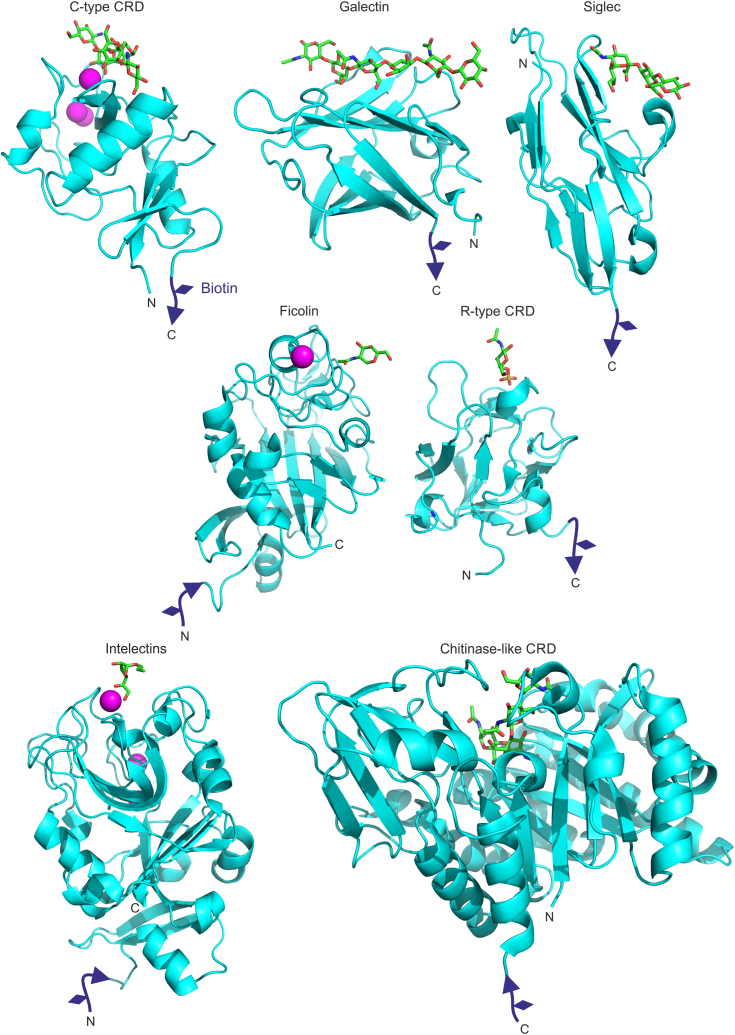
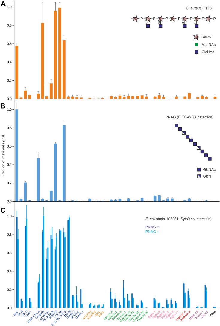
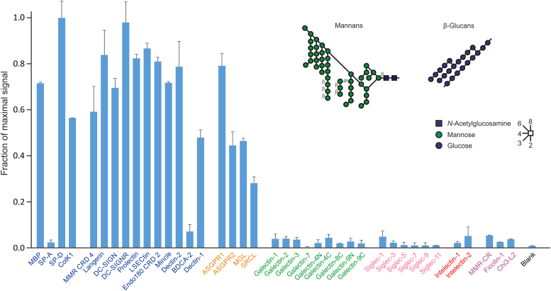
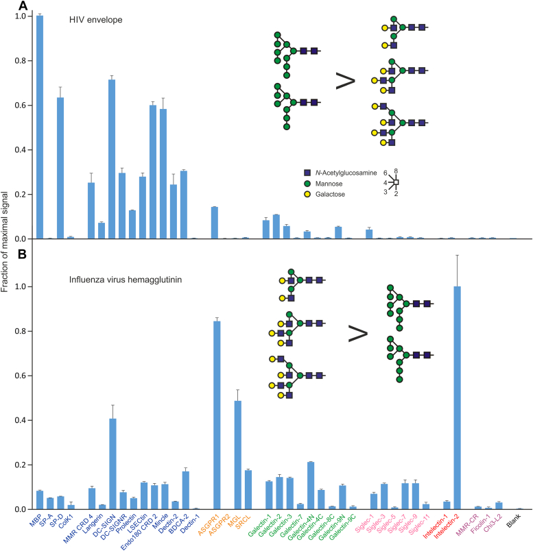
A human lectin array has been developed to probe the interactions of innate immune receptors with pathogenic and commensal microorganisms. Following the successful introduction of a lectin array containing all of the cow C-type carbohydrate-recognition domains (CRDs), a human array described here contains the C-type CRDs as well as CRDs from other classes of sugar-binding receptors, including galectins, siglecs, R-type CRDs, ficolins, intelectins, and chitinase-like lectins. The array is constructed with CRDs modified with single-site biotin tags, ensuring that the sugar-binding sites in CRDs are displayed on a streptavidin-coated surface in a defined orientation and are accessible to the surfaces of microbes. A common approach used for expression and display of CRDs from all of the different structural categories of glycan-binding receptors allows comparisons across lectin families. In addition to previously documented protocols for binding of fluorescently labeled bacteria, methods have been developed for detecting unlabeled bacteria bound to the array by counter-staining with DNA-binding dye. Screening has also been undertaken with viral glycoproteins and bacterial and fungal polysaccharides. The array provides an unbiased screen for sugar ligands that interact with receptors and many show binding not anticipated from earlier studies. For example, some of the galectins bind with high affinity to bacterial glycans that lack lactose or N-acetyllactosamine. The results demonstrate the utility of the human lectin array for providing a unique overview of the interactions of multiple classes of glycan-binding proteins in the innate immune system with different types of microorganisms.

Keywords: array screening; carbohydrate-binding protein; glycan-binding receptors; glycobiology; host-pathogen interaction; innate immunity; lectin.


Conflict of interest statement

Conflict of interest The authors declare that they have no conflicts of interest with the contents of this article.

Figures

Figure 1
Figure 1
Attachment of biotin tags to CRDs from different structural groups. Representative structures for each group are shown: C-type CRD from DC-SIGN with Man3GlcNAc2 oligosaccharide (PDB 1K9I); galectin-9 with N-acetyllactosamine trimer (PDB 2ZHM); siglec 5 CRD with 3′-sialyllactose (PDB 2ZG3); ficolin 1 CRD with GalNAc (PDB 2JHI); R-type CRD from the mannose receptor with 4-sulfo-N-acetylgalactosamine (PDB 1DQO); intelectin 1 with allyl-β-galactofuranose (PDB 4WMY); and chitinase-like lectin Chi3L2 with (GlcNAcβ1-4)4 oligosaccharide (PDB 4P8W). Figure was generated with PyMol based on the indicated Protein Data Bank (PDB) files. Sugar ligands are shown as sticks, with carbon atoms in green, oxygen atoms in red, and nitrogen atoms in blue. Ca2+ is shown as magenta spheres and the appended biotin tags are in purple. CRD, carbohydrate-recognition domain; PDB, Protein Data Bank.
Figure 2
Figure 2
Interaction of Klebsiella pneumoniae with lectins on the array. Human lectin array was screened with versions of two strains of K. pneumoniae with and without capsules. Cells expressing GFP were grown to stationary phase and fixed with paraformaldehyde. Results were normalized to the maximum signal for the unencapsulated strain in each panel. For each strain, a representative example from three experiments is shown. A, screening with strain B5055, serotype K2:O1v1, and a mutant lacking the capsule at concentrations of 2.5 to 5 × 108 cells/ml. The structure of the O1v1 outer polysaccharide is shown. Average errors were 12%. B, screening with strain ICC8001, serotype K2:O1v2, and mutants lacking either the capsule or the O-antigen at concentrations of 5 to 10 × 108 cells/ml. The structure of the O1v2 outer polysaccharide is shown. Average errors were 10%. Data are reported in Table S1.
Figure 3
Figure 3
Enteropathogenic Escherichia coli binding to the human lectin array. Enteropathogenic E. coli strain E2348/69 (O127:H6) grown to stationary phase and fixed with paraformaldehyde were used to screen the array. Cells expressing GFP were screened at 1.6 × 108 cells/ml (low concentration) and 8 × 108 cells/ml (high concentration). Cells without GFP were screened at 16 × 108 cells/ml and were visualized by counter-staining with Syto 9 dye. Average errors were 10%, 9%, and 3% for the three different protocols. A representative example from two experiments is shown. Results for each experiment were normalized to the highest signal. Structure of two repeat units from the O127 outer polysaccharide of lipopolysaccharide is shown. Data are reported in Table S2.
Figure 4
Figure 4
Screening of human lectin array with Staphylococcus aureus and PNAG.A, screening with FITC-labeled, heat-killed cells of the Wood 46 strain of S. aureus at a concentration of 1 × 108 cells/ml. Average errors were 13%. B, screening with PNAG at 60 μg/ml, followed by visualization with FITC-labeled wheat germ agglutinin at 40 μg/ml. Average errors were 7%. C, screening with Escherichia coli strain E. coli JC8031 and a mutant lacking PNAG at 4 to 12 × 108 cells/ml. Bound cells were counterstained with Syto 9 dye. Average errors were 10%. Results were normalized to the highest signal in each experiment. Structures of wall teichoic acid from S. aureus, showing α-linked GlcNAc residues, and of PNAG are shown. A representative example from two experiments is shown. Data are reported in Table S3. PNAG, poly-N-acetylglucosamine.
Figure 5
Figure 5
Binding of yeast zymosan to human lectin array. FITC-labeled zymosan was used for screening at a concentration of 1 × 107 particles/ml. Average errors were 7%. A representative example from two experiments is shown. Structures of the predominant yeast wall mannans and β-glucans in zymosan are shown. Ligands for galactose-binding receptors are also present, but the relevant structures are not known (66). Data are reported in Table S4.
Figure 6
Figure 6
Viral glycoproteins tested on human lectin array. Soluble, trimeric forms of the surface glycoproteins were generated using trimerization sequences to replace the membrane anchors. Purified proteins were labeled with Alexa488 and used to screen the array. A, HIV BG505 SOSIP.664 Envelope glycoprotein (7.9 μg/ml). Average errors were 6%. B, influenza virus H3 Brisbane/2007 hemagglutinin (6.2 μg/ml). Average errors were 7%. For each protein, a representative example from three experiments, covering a 10-fold concentration range, is shown. Examples of potential target glycans are shown. Glycans present on HIV envelope protein are predominantly high mannose N-linked oligosaccharides with up to nine mannose residues. More glycans on influenza virus hemagglutinin are complex glycans, with variable numbers of branches, often terminating in galactose. Data are reported in Table S5.

References

    1. Drickamer K., Taylor M.E. Recent insights into structures and functions of C-type lectins in the immune system. Curr. Opin. Struct. Biol. 2015;34:26–34. - PMC - PubMed
    1. Taylor M.E., Drickamer K. Mammalian sugar-binding receptors: known functions and unexplored roles. FEBS J. 2019;286:1800–1814. - PMC - PubMed
    1. Rillahan C.D., Paulson J.C. Glycan microarrays for decoding the glycome. Annu. Rev. Biochem. 2018;80:797–823. - PMC - PubMed
    1. Li Z., Feizi T. The neoglycolipid (NGL) technology-based microarrays and future prospects. FEBS Lett. 2018;592:3976–3991. - PubMed
    1. Stowell S.R., Arthur C.M., McBride R., Berger O., Razi N., Heimburg-Molinaro J., et al. Microbial glycan microarrays define key features of host-microbial interactions. Nat. Chem. Biol. 2014;10:470–476. - PMC - PubMed

Publication types

LinkOut - more resources