. 2024 Dec 1;86(12):1273-1278.
doi: 10.1292/jvms.24-0338. Epub 2024 Oct 9.

Attenuation of the neuropathogenic equine herpesvirus type 1 strain Ab4p in hamsters by a single amino acid mutation (D752N) in viral DNA polymerase ORF30

Affiliations

Attenuation of the neuropathogenic equine herpesvirus type 1 strain Ab4p in hamsters by a single amino acid mutation (D752N) in viral DNA polymerase ORF30

Fuka Nishimura et al. J Vet Med Sci. .

Abstract

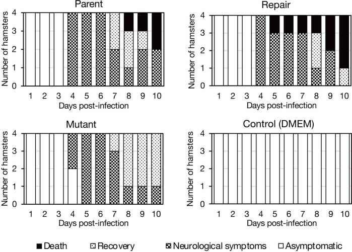
Equine herpesvirus type 1 (EHV-1) causes abortion, respiratory infection, and neurological diseases (equine herpesvirus myeloencephalopathy) in horses. A single nucleotide polymorphism (SNP) associated with a single amino acid in the DNA polymerase gene (ORF30, in which D752 is neuropathogenic and N752 is non-neuropathogenic) of EHV-1 has been associated with neuropathogenicity in horses. We constructed an EHV-1 Ab4p ORF30 N752 mutant and a repair virus to examine the effect of a D752N mutation on the neuropathogenicity of the virus in Syrian hamsters. The N752 mutation did not affect viral growth in cultured cells but it did attenuate the neuropathogenicity of Ab4p in the hamsters. The results suggest that D752N is involved in neuropathogenicity not only in horses but also in hamsters.

Keywords: DNA polymerase; ORF30; a single nucleotide polymorphism (SNP); equine herpesvirus; equine herpesvirus encephalopathy (EHM).


Conflict of interest statement

The authors have nothing to disclose.

Figures

Fig. 1.
Fig. 1.
Method for producing pAb4p ORF30 A2254 mutant bacterial artificial chromosome (BAC). The figure on top shows the pAb4p BAC before modification and where it is modified. pZC320 is a low-copy-number cloning vector derived from plasmid F [29]. The BAC was modified in the following five steps (shown as (1), (2)) in the figure: (1) A 1,539 bp fragment of ORF30 from base number 1,573 to 3,080 containing G2254 was amplified by PCR using primers Infusion ORF30 pUC19 F and R, which include 16 bases and 15 bases, respectively, of the homologous sequence of pUC19. (2) The ORF30 fragment was linked to pUC19 for constructing pUC-ORF30 plasmid. (3) G2254 was converted to A2254 by inverse PCR using primers ORF30 G2254A F and R and PrimeSTAR mutagenesis kit. (4) A rpsL-neo cassette (1,418 bp), which was amplified using primers Δ-30 rpsL-neo F and R and a 1,319 bp synthetic rpsL-neo fragment as a template, was introduced into E. coli harboring pAb4p BAC 2019 and the pRedET plasmid. The rpsL-neo cassette is shown as a gray rectangle in Fig. 1. A region from 1,731 to 3,049 including G2254 in ORF30 was replaced by the rpsL-neo cassette, resulting in pAb4p∆ORF30rpsLneo BAC plasmid. (5) A 1,476 bp fragment corresponding to base numbers 1,605 to 3,080 of ORF30 was amplified by PCR using a pair of primers, ΔrpsL ORF30F and R, and pUC19-ORF30 plasmid which was constructed in the step (3) as a template. The rpsL-neo cassette in pAb4p∆ORF30 rpsLneo was replaced by the fragment using Red/ET recombination, resulting in the completed pAb4p ORF30 G2254 bacterial artificial chromosome (BAC) plasmid.
Fig. 2.
Fig. 2.
Restriction enzyme-treated fragments electrophoresis images of viral BAC DNA from the parental strain, the G2254A mutant, and the repair. In the parental strain and mutant, the second and third bands from the top were identified side by side, whereas in the G2254A mutant, only one band was identified in the second band from the top (arrow). 1: 1kb ladder, 2: λ DNA HindIII digest, 4-6: Sal I treated samples, 8-10: EcoR I treated samples, 12-14: HindIII treated samples. Within each group of restriction enzyme-treated samples, from left to right: parental strain, G2254A mutant, repair.
Fig. 3.
Fig. 3.
Multistep growth experiments in FHK Tcl 3.1 cells. FHK Tcl 3.1 cells were inoculated with virus at multiplicity of infection (m.o.i.)=0.01. Supernatants were collected at 0, 24, 48, and 72 hr after inoculation and stored at −80°C before titer assays were performed on the FHK Tcl3.1 cells. Differences in virus titers were statistically analyzed using the Tukey-Kramer method using RStudio 2024.04.2+764. Mean differences were considered significant when the P value was less than 0.05.
Fig. 4.
Fig. 4.
Body weight change of virus-inoculated Syrian hamsters. Four animals per group were inoculated intranasally with the virus at a dose of 6 × 10³ pfu per animal and observed for 10 days for changes in body weight and the presence of neurological symptoms. The graph shows the weight change with the inoculation date as 1.0. Group means and standard deviations are shown in graphs. Dagger (†) indicates that the hamster died. Dunnet’s test was performed using RStudio 2024.04.2+764. Asterisk (*) indicates statistically significant differences: P<0.05, comparing to the control group (DMEM). The numbers indicate the cumulative number of dead hamsters.
Fig. 5.
Fig. 5.
Neurological symptoms of virus-inoculated Syrian hamsters. Par.: parental inoculation group; Mut.: mutant inoculation group; Rev.: repair inoculation group; DMEM: DMEM inoculation group. Black indicates death (R3 only, day 5) or reaching the humane endpoint (other); Light hatch indicates recovery; dark hatch indicates neurological symptoms. Blank cells indicate that no symptoms were observed.
Fig. 6.
Fig. 6.
Histopathology of the dead hamster brains. Coarsening in the piriform lobes was observed in each of the parent (A), repair (B), and mutant (C) virus inoculated hamster brains. No lesions were observed in the piriform lobe of control hamster brains (D). Bar, 100 µm.

Similar articles

References

    1. Awan AR, Chong YC, Field HJ. 1990. The pathogenesis of equine herpesvirus type 1 in the mouse: a new model for studying host responses to the infection. J Gen Virol 71: 1131–1140. doi: 10.1099/0022-1317-71-5-1131 - DOI - PubMed
    1. Azmi M, Field HJ. 1993. Interactions between equine herpesvirus type 1 and equine herpesvirus type 4: T cell responses in a murine infection model. J Gen Virol 74: 2339–2345. doi: 10.1099/0022-1317-74-11-2339 - DOI - PubMed
    1. Chong YC, Duffus WP. 1992. Immune responses of specific pathogen free foals to EHV-1 infection. Vet Microbiol 32: 215–228. doi: 10.1016/0378-1135(92)90146-K - DOI - PubMed
    1. Field HJ, Awan AR, de la Fuente R. 1992. Reinfection and reactivation of equine herpesvirus-1 in the mouse. Arch Virol 123: 409–419. doi: 10.1007/BF01317274 - DOI - PubMed
    1. Frampton AR, Jr, Smith PM, Zhang Y, Grafton WD, Matsumura T, Osterrieder N, O’Callaghan DJ. 2004. Meningoencephalitis in mice infected with an equine herpesvirus 1 strain KyA recombinant expressing glycoprotein I and glycoprotein E. Virus Genes 29: 9–17. doi: 10.1023/B:VIRU.0000032785.19420.14 - DOI - PubMed