. 2024 Oct;21(6):e00458.
doi: 10.1016/j.neurot.2024.e00458. Epub 2024 Oct 8.

Targeting the Hippo pathway in Schwann cells ameliorates peripheral nerve degeneration via a polypharmacological mechanism

Affiliations

Targeting the Hippo pathway in Schwann cells ameliorates peripheral nerve degeneration via a polypharmacological mechanism

Hyung-Joo Chung et al. Neurotherapeutics. 2024 Oct.

Abstract

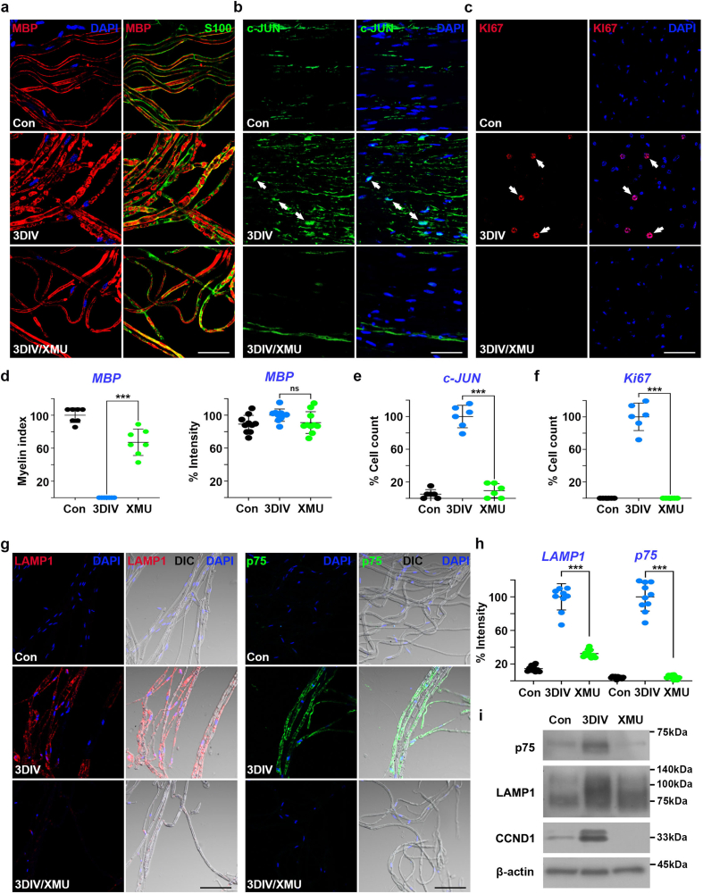
Peripheral neuropathies (PNs) are common diseases in elderly individuals characterized by Schwann cell (SC) dysfunction and irreversible Wallerian degeneration (WD). Although the molecular mechanisms of PN onset and progression have been widely studied, therapeutic opportunities remain limited. In this study, we investigated the pharmacological inhibition of Mammalian Ste20-like kinase 1/2 (MST1/2) by using its chemical inhibitor, XMU-MP-1 (XMU), against WD. XMU treatment suppressed the proliferation, dedifferentiation, and demyelination of SCs in models of WD in vitro, in vivo, and ex vivo. As a downstream mediator of canonical and noncanonical Hippo/MST1 pathway activation, the mature microRNA (miRNA) let-7b and its binding partners quaking homolog (QKI)/nucleolin (NCL) modulated miRNA-mediated silencing of genes involved in protein transport. Hence, direct phosphorylation of QKI and NCL by MST1 might be critical for WD onset and pathogenesis. Moreover, p38α/mitogen-activated protein kinase 14 (p38α) showed a strong affinity for XMU, and therefore, it may be an alternative XMU target for controlling WD in SCs. Taken together, our findings provide new insights into the Hippo/MST pathway function in PNs and suggest that XMU is a novel multitargeted therapeutic for elderly individuals with PNs.

Keywords: Hippo/MST pathway; RNA-binding protein; Schwann cells; let-7b; p38α/MAPK14.


Conflict of interest statement

Declaration of competing interest The authors declare that they have no known competing financial interests or personal relationships that could have appeared to influence the work reported in this paper.

Figures

Fig. 1
Fig. 1
XMU inhibits peripheral nerve degeneration. a, b Microscopy of the transverse stripes (a, Scale bar ​= ​1 ​mm) and ovoid fragments (b, Scale bar ​= ​50 ​μm) from the mouse sciatic nerve explants at three days in vitro (3DIV) in the presence or absence of XMU (100 ​μM). Control (Con) sample indicates the sciatic nerves right after explantation. Arrows (b) indicated the ovoids (myelin fragments). c, d Number of the stripes (c) and the ovoids (d) in a and b, respectively, were measured. e Transgenic adult zebrafish [Tg(MBP:EGFP) was used as in vivo WD model. Fluorescence images of zebrafish caudal fin were acquired at 10 days post-injury (DPI) with or without XMU treatment (10 ​μM) under a fluorescence stereoscope. Scale bar ​= ​1 ​mm. f, g ​The number of intact GFP fluorescence (MBP nerves, ​f) and its intensity (MBP intensity, g) were measured at three areas (1×1 mm2). h-j Expression patterns of MST1, phosphorylated MST1 at T183 residue (p-MST1), and YAP in ex vivo sciatic nerves at 3DIV in the presence or absence of XMU (100 ​μM). Scale bar ​= ​50 ​μm. MST1 (h), p-MST1 (i), or YAP (j) were co-stained with either S100 (green, a marker of SCs) or 4′,6-Diamidine-2′-phenylindole (DAPI, blue). Dotted boxes indicated higher magnification images of the nucleus in Schwann cells (Scale bar ​= ​20 ​μm). k, l Graphs showed the morphometric results of MST1 (% intensity, k) and p-MST (% intensity, l) in h and i, respectively. m Graphs showed the morphometric results of YAP (% cell counts) in (j). n Western blotting of hTERT02.3 ​cell lysates in the presence or absence of XMU (10 ​μM) showed the expression patterns of MST1, LATS1, phosphorylated LATS1 (p-LATS1), and YAP as components of canonical Hippo pathways in human SCs. ∗p ​< ​0.05, ∗∗p ​< ​0.01, ∗∗∗p ​< ​0.001, ns: not significant.
Fig. 2
Fig. 2
XMU ameliorates degenerative characteristics in SCs. a-c Expression patterns of markers of demyelination (MBP, red; a), negative regulator for myelination (c-JUN, green; b), and proliferation (Ki-67, red; c) in ex vivo sciatic nerve fibers at 3DIV in the presence or absence of XMU (100 ​μM). S100 (green) and DAPI (blue) were co-stained with each marker. Control (Con) was non-injured sciatic nerves. The teasing samples, longitudinal sections, and cross-sections of the sciatic nerve were used for staining MBP, c-JUN, and KI-67, respectively. Scale bar ​= ​50 ​μm. d Myelin index was the number of consecutive double-lines of MBP in each sample. Percent intensity of MBP signals was quantified by counting the relative number of pixels showing each intensity. e, f Percentage of cell counts indicated the number of c-JUN/DAPI (e) or KI-67/DAPI (f) double-positive nuclei in each sample. g Immunofluorescence in sciatic nerve fibers stained with lysosomal-associated membrane protein 1 (LAMP1, red) or p75 neurotrophin receptor (p75, green) as markers of SC dedifferentiation and DAPI (blue). Scale bar ​= ​50 ​μm. h The graph showed the intensity of LAMP1 and p75 staining in sciatic nerve fibers cultured with 100 ​μM of XMU for 3DIV. i Western blot analysis of p75, LAMP1, and cyclin D1 (CCND1, a cell proliferation marker) levels in ex vivo sciatic nerve lysates. β-actin is a loading control. ∗∗∗p ​< ​0.001, ns: not significant.
Fig. 3
Fig. 3
XMU alters protein expression in dedifferentiated SCs. a. Gene Ontology (GO) analysis was performed on upregulated proteins in XMU-treated SW10 ​cells compared with control cells using a threefold (log2 value) upregulated cutoff. A GO scatter plot was shown using Reduce and Visualize Gene Ontology (REVIGO). GO terms are represented by the bubbles, and the colors are meant as the P values. b. Representative GO terms of group 1 were shown, and they were selected among the phenotypic GO terms in Schwann cells. c. XMU-dependent genes involved in protein transport are validated via qRT-PCR. The nine genes were downregulated at 3DIV. Mouse sciatic nerve explants were used for the validation. ∗p ​< ​0.05, ∗∗p ​< ​0.01, and ∗∗∗p ​< ​0.001. d. Venn diagram showing the comparison of 484 genes associated with neuropathy (NP), and proteins from global proteomics. Proteins for NP (UMLS CUI#C0442874) were collected from the DisGeNET (v7.0) database. Transcription factor, TF. e. GO terms analysis by using the 181 proteins. f. 12 shared transcription factors are represented in the black square boxes. The expression of p-c-JUN was validated by Western blot analysis in SW10 ​cells. g. Functional protein association network of genes shown in key transcription factors and genes involving in protein transport. The size of the circle represents the STRING interaction score.
Fig. 4
Fig. 4
Mature miRNA let-7b mediates MST1-dependent WD. aEx vivo FISH showing let-7b enrichment and subcellular location in sciatic nerve fibers using anti-let-7b-Cy3 DNA (let-7b probe) in the presence or absence of XMU. Scale bar ​= ​50 ​μm. miRNA-FISH (red) was co-stained with either S100 (green) or DAPI (blue). DNA [(-)DNA, TTGACAGGCAACCCCAACCA] template was used as a negative control. Arrows and arrowheads indicated anti-let-7b/DAPI and anti-let-7b/S100 double-positive signals, respectively. Blank arrows also indicated the nuclei with no signals of anti-let-7b-Cy3. b Sciatic nerve explants transfected with let-7b (1 ​μM) or anti-let-7b (1 ​μM) labeled with Cy3 were observed via confocal microscope at 3DIV. Scale bar ​= ​200 ​μm. Transfected nerve fibers were shown as red (let-7b) and green (anti-let-7b). c Transverse stripes in let-7b-Cy3 (RNA)- or anti-let-7b-Cy3 (DNA)-transfected ex vivo samples at 3DIV. RNA [(-)RNA, GCGCGCUUUGUAGGAUUCG] or (-)DNA (a) templates were used as negative controls. Graphs displayed the stripe number of each group. Dotted boxes indicated transverse stripes. Scale bar ​= ​1 ​mm. d Differential interference contrast (DIC) or fluorescence images of ovoid fragments in let-7b-Cy3 (RNA)- or anti-let-7b-Cy3 (DNA)-transfected ex vivo samples at 3DIV. Black arrows indicated Cy3-labeled nucleotides-transfected nerve fibers protected from WD. The graph showed the ovoid numbers of each group. Scale bar ​= ​50 ​μm. e qRT-PCR validation of let-7b and let-7c in ex vivo sciatic explants. fSnap23 and Tram2, genes involved in protein transport, were validated via qRT-PCR in ex vivo sciatic explants. ∗ or #p ​< ​0.05, ∗∗p ​< ​0.01, and ∗∗∗p ​< ​0.001, ns: not significant.
Fig. 5
Fig. 5
MST1 phosphorylates QKI and NCL to modulate let-7b level in SCs. a Expression pattern of QKI5 in ex vivo nerve fibers with or without XMU treatment at 3DIV was assessed by immunostaining analysis. Arrows indicated QKI5/DAPI double-positive signals. Scale bar ​= ​50 ​μm. b Immunostaining of NCL in ex vivo sciatic nerve fibers with or without XMU treatment at 3DIV. White arrows indicated the number of NCL/DAPI double-positive signals in each sample. Blank arrows showed NCL located around the nuclei, not in the nuclei. Scale bar ​= ​50 ​μm. c, d Percentage of cell counts indicated the number of QKI5/DAPI (c) or NCL/DAPI (d) double-positive signals. e Molecular docking model of human QKI bound with a fragment of let-7b, UGUGGU (left panel). Surface Plasmon Resonance (SPR) profiles of recombinant human QKI proteins and let-7b with biotin label (right panel). f Crystal structure (left panel) of mouse NCL bound to NCL recognition element (NRE). The phosphorylated threonines are located outside of 306–470 a.a. range. Fluorescence polarization of let-7b-Cy3 bound to recombinant mouse NCL protein (right panel). Quaking, QKI; nucleolin, NCL. g, h At 48 ​h after transfection of empty vector or QKI/NCL plasmids, the lysates from SW10 ​cells were subjected to SDS-PAGE for Western blot analysis against QKI (g) or NCL (h) (left panel). Genes involving protein transport in total RNA from the transfected SW10 ​cells were detected by qRT-PCR (right panel). The 9 genes were downregulated after QKI/NCL overexpression. n ​= ​3. i, j At 48 ​h after transfection of control and QKI/NCL shRNA in SW10, QKI (i) or NCL (j) from cell lysates were detected by Western blot (left panel). The genes involving protein transport were detected by qRT-PCR (right panel). The 9 genes were upregulated after QKI/NCL knockdown. n ​= ​3. ∗p ​< ​0.05, ∗∗p ​< ​0.01, and ∗∗∗p ​< ​0.001.
Fig. 6
Fig. 6
XMU interacts with p38α protein kinase as its novel drug target in SCs. a Kinase-substrate enrichment analysis without phosphatases. Proteome normalized ratio of XMU/DMSO was taken to infer Z-score (kinase activation score). Red or blue presented kinases significantly activated and attenuated, respectively. b Binding affinity of XMU with the highly ranked 12 protein kinase targets. Affinity was defined as kcal/mol. p38α showed the strongest binding with XMU among the 12 kinases in a. c, d Expression patterns of p38α (c) and p-p38α (d) in ex vivo nerve fibers with or without XMU treatment at 3DIV were assessed by immunostaining analysis. Arrows indicated p38α or p-p38α/DAPI double-positive signals. Scale bar ​= ​50 ​μm. e Western blot analysis of p38α was performed by using sciatic nerve explants. Graphs showed the intensities of immunoblots in each sample. f The left graph showed the intensities of p38α staining in sciatic nerve fibers cultured with 100 ​μM of XMU for 3DIV in c. Percentage of cell counts (right panel) indicated the number of p-p38α/DAPI-positive signals in d. g DARTS assay for target validation. Thermolysin-dependent p38α protein destabilization was suppressed by treatment and binding of XMU in SW10 ​cells. The graph (lower panel) is the quantification data shown in the Western blot (upper panel). h Schematic diagram of the polypharmacological mechanism of XMU. Back and brown arrows implied XMU-dependent canonical and non-canonical pathways, respectively, in SCs during WD. ∗∗∗p ​< ​0.001.

References

    1. Waller A. Experiments on the section of the glosso-pharyngeal and hypoglossal nerves of the frog, and observations of the alterations produced thereby in the structure of their primitive fibres. Edinb Med Surg J. 1851;76:369–376. - PMC - PubMed
    1. Stoll G., Jander S., Myers R.R. Degeneration and regeneration of the peripheral nervous system: from Augustus Waller's observations to neuroinflammation. J Peripher Nerv Syst. 2002;7:13–27. - PubMed
    1. Suzuki M. Peripheral neuropathy in the elderly. Handb Clin Neurol. 2013;115:803–813. - PubMed
    1. Brisset M., Nicola G. Peripheral neuropathies and aging. Geriatr Psychol Neuropsychiatr Vieil. 2018;16:409–413. - PubMed
    1. Tricaud N., Park H.T. Wallerian demyelination: chronicle of a cellular cataclysm. Cell Mol Life Sci. 2017;74:4049–4057. - PMC - PubMed

MeSH terms

LinkOut - more resources