Observational Study
. 2024 Oct 9;14(1):23629.
doi: 10.1038/s41598-024-74088-y.

Adaptive optics scanning laser ophthalmoscopy in a heterogenous cohort with Stargardt disease

Affiliations
Observational Study

Adaptive optics scanning laser ophthalmoscopy in a heterogenous cohort with Stargardt disease

Mital Shah et al. Sci Rep. .

Abstract

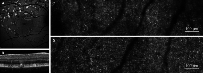
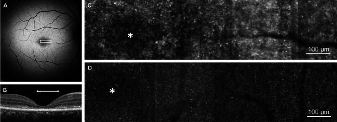
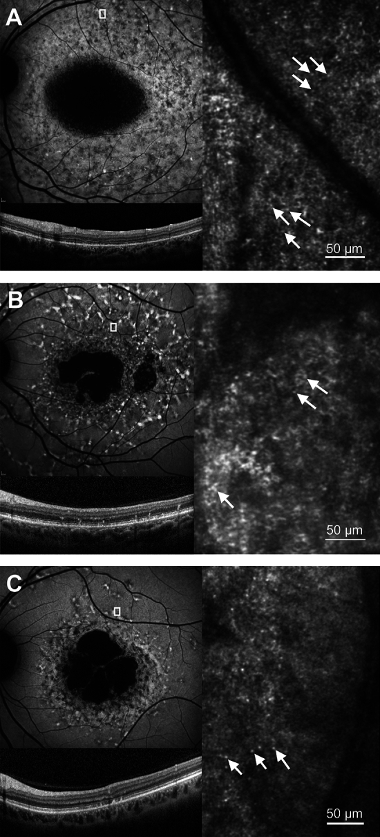
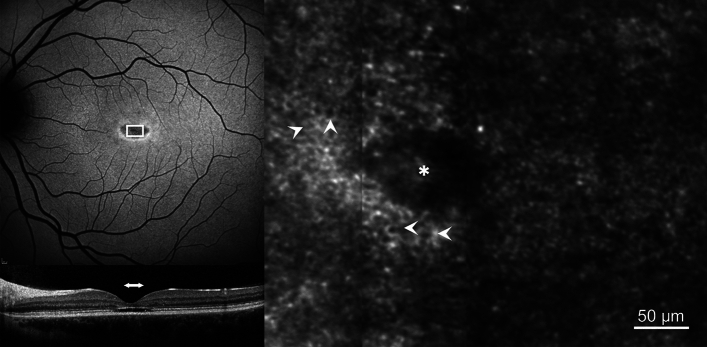
Image based cell-specific biomarkers will play an important role in monitoring treatment outcomes of novel therapies in patients with Stargardt (STGD1) disease and may provide information on the exact mechanism of retinal degeneration. This study reports retinal image features from conventional clinical imaging and from corresponding high-resolution imaging with a confocal adaptive optics scanning laser ophthalmoscope (AOSLO) in a heterogenous cohort of patients with Stargardt (STGD1) disease. This is a prospective observational study in which 16 participants with clinically and molecularly confirmed STGD1, and 7 healthy controls underwent clinical assessment and confocal AOSLO imaging. Clinical assessment included short-wavelength and near-infrared fundus autofluorescence, spectral-domain optical coherence tomography, and macular microperimetry. AOSLO images were acquired over a range of retinal eccentricities (0°-20°) and mapped to areas of interest from the clinical images. A regular photoreceptor mosaic was identified in areas of normal or near normal retinal structure on clinical images. Where clinical imaging indicated areas of retinal degeneration, the photoreceptor mosaic was disorganised and lacked unambiguous cones. Discrete hyper-reflective foci were identified in 9 participants with STGD1 within areas of retinal degeneration. A continuous RPE cell mosaic at the fovea was identified in one participant with an optical gap phenotype. The clinical heterogeneity observed in STGD1 is reflected in the findings on confocal AOSLO imaging.

Keywords: ABCA4; AOSLO; Adaptive optics; Adaptive optics scanning laser ophthalmoscope; Retinal imaging; Stargardt disease.


Conflict of interest statement

The authors declare no competing interests.

Figures

Fig. 1
Fig. 1
Photoreceptor mosaic in Stargardt disease in an area of near normal retina. (A) 30° short-wavelength fundus autofluorescence image, white box represents the retinal area corresponding to the photoreceptor mosaic in C and retinal cross section in B. (B) Spectral-domain optical coherence tomography of the retinal region represented by the white box in A. (C) Photoreceptor mosaic of participant S04. (D) Photoreceptor mosaic of an age-matched control (C05) to participant S04 from the same retinal eccentricity.
Fig. 2
Fig. 2
Photoreceptor mosaic in Stargardt disease in an area of degenerated retina. (A) 30° short-wavelength fundus autofluorescence image, white box represents the retinal area corresponding to the photoreceptor mosaic in C. (B) Spectral-domain optical coherence tomography. The horizontal white arrow indicates the retinal area that corresponds to the white box in A. (C) Photoreceptor mosaic of participant S06. (D) Photoreceptor mosaic of an age-matched control (C02) to participant S06 from the same retinal eccentricity. Asterisk represents the foveal centre.
Fig. 3
Fig. 3
The relationship between retinal eccentricity and discrete hyper-reflective foci size. Each datum represents the size of an averaged image stack of 200 discrete hyper-reflective foci taken from a single AOSLO image. This is reported as full width at half maximum of a Gaussian profile fit to the average image. WDHF—full width at half maximum size of the discrete hyper-reflective foci in microns. The mean size of the discrete hyper-reflective foci for all participants and all retinal eccentricities is 4.1 μm (SD = 0.5 μm). The solid line represents a linear least-squares regression line of best fit (r = − 0.47, p < 0.001).
Fig. 4
Fig. 4
Examples of discrete hyper-reflective foci. 30° short-wavelength fundus autofluorescence (upper left), spectral-domain optical coherence tomography (lower left) and confocal AOSLO (right) images are shown. The white box in the short- wavelength fundus autofluorescence image represents the retinal area corresponding to the photoreceptor mosaic. White arrows indicate examples of the discrete hyper-reflective foci that can be seen throughout each image. (A) participant S11, (B) participant S14, (C) participant S15.
Fig. 5
Fig. 5
Retinal pigment epithelial (RPE) mosaic. 30° short-wavelength fundus autofluorescence (SW-FAF, upper left), spectral-domain optical coherence tomography (SD-OCT, lower left) and confocal AOSLO (right) images are shown. The white box in the SW-FAF image represents the retinal area corresponding to the photoreceptor mosaic. The horizontal white arrow in the SD-OCT image indicates the retinal area that corresponds to the white box in the SW-FAF image. The white arrowheads in the confocal AOSLO image indicate examples of the RPE cells visible throughout the image. Asterisk represents the foveal centre.

References

    1. Tanna, P., Strauss, R. W., Fujinami, K. & Michaelides, M. Stargardt disease: Clinical features, molecular genetics, animal models and therapeutic options. Br. J. Ophthalmol.101, 25–30 (2017). - PMC - PubMed
    1. Spiteri Cornish, K. et al. The epidemiology of Stargardt disease in the United Kingdom. Ophthalmol. Retin.1, 508–513 (2017). - PubMed
    1. Al-Khuzaei, S. et al. An overview of the genetics of ABCA4 retinopathies, an evolving story. Genes12, 1241 (2021). - PMC - PubMed
    1. Al-Khuzaei, S. et al. The role of multimodal imaging and vision function testing in ABCA4-related retinopathies and their relevance to future therapeutic interventions. Ther. Adv. Ophthalmol.13, 25158414211056384 (2021). - PMC - PubMed
    1. Schulz, H. L. et al. Mutation spectrum of the ABCA4 gene in 335 stargardt disease patients from a multicenter German cohort—impact of selected deep intronic variants and common SNPs. Investig. Ophthalmol. Vis. Sci.58, 394–403 (2017). - PMC - PubMed

Publication types

LinkOut - more resources