

Low-frequency rTMS Plays a Neuroprotective role in Pilocarpine-induced Status Epilepticus Rat Models Through the AMPAR GluA1-STIM-Ca2+ Pathway
- PMID: 39384697
- PMCID: PMC11880165
- DOI: 10.1007/s12035-024-04521-w
Low-frequency rTMS Plays a Neuroprotective role in Pilocarpine-induced Status Epilepticus Rat Models Through the AMPAR GluA1-STIM-Ca2+ Pathway
Abstract
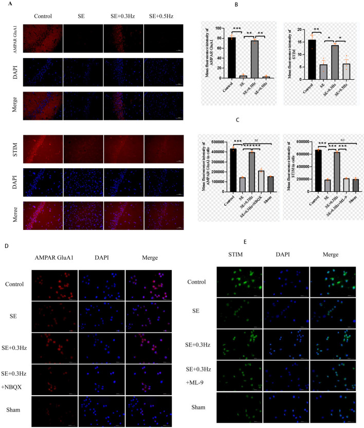
Low-frequency repetitive transcranial magnetic stimulation (rTMS) refers to the stimulation of the brain using repetitive magnetic field pulses at a low frequency (≤ 1 Hz) to reduce seizures. Currently, the mechanism is not well understood. Male Sprague-Dawley rats underwent pilocarpine-induced status epilepticus (SE) and were then stimulated with low-frequency rTMS. An epilepsy cell model was then established by incubating rat hippocampal neurons with Mg2+-free extracellular fluids. The effects of the low-frequency rTMS on epileptogenesis and hippocampal neuron injury were evaluated using a video electroencephalogram (vEEG) and Nissl staining, and the expression of AMPAR GluA1 and STIM in the hippocampus and hippocampal neurons was assessed using western blot and immunofluorescence. Additionally, the intracellular Ca2+ concentration and reactive oxygen species (ROS) were measured using flow cytometry. Low-frequency rTMS attenuated spontaneous recurrent seizures in rats with epilepsy, with the SE group exhibiting a higher incidence (100%) and frequency (3.00 ± 0.18 times/day) than the SE + 0.3 (50% incidence, 0.06 ± 0.03 times/day), SE + 0.5 (0.20 ± 0.02 times/day) and SE + 1 Hz (1.02 ± 0.05 times/day) groups. Additionally, rTMS reduced the damage and apoptosis of hippocampal pyramidal neurons, increasing their numbers in the CA1 and CA3 regions. Furthermore, AMPAR GluA1 and STIM expression were upregulated in the hippocampus when using rTMS, reversing the downregulation caused by seizures. Immunofluorescence verified the increased fluorescence intensity of AMPAR GluA1 and STIM. Moreover, rTMS inhibited Ca2+ overload and ROS in epileptic neuron models. Low-frequency rTMS may exert neuroprotective effects through the AMPAR GluA1-STIM-Ca2+ pathway.
Keywords: Epilepsy cell Model; Low-frequency Repetitive Transcranial Magnetic Stimulation; Neuroprotection; Temporal lobe Epilepsy.
© 2024. The Author(s).
Conflict of interest statement
Declarations. Consent for Publication: Not applicable. Competing Interests: The authors declare no competing interests. Ethics Approval: The animal study was reviewed and approved by the Second Hospital of Hebei Medical University (2022-AE004).
Figures


References
-
- Reddy DS, Golub VM, Ramakrishnan S, Abeygunaratne H, Dowell S, Wu X (2022J) A Comprehensive and Advanced Mouse Model of Post-Traumatic Epilepsy with Robust Spontaneous Recurrent Seizures. Curr Protoc. 2(6)e447. 10.1002/cpz1.447 - PubMed
-
- Trinka E, Cock H, Hesdorffer D, Rossetti AO, Scheffer IE, Shinnar S, Shorvon S, Lowenstein DH (2015) A definition and classification of status epilepticus–Report of the ILAE Task Force on Classification of Status Epilepticus. Epilepsia. 56(10):1515–23. 10.1111/epi.13121 - PubMed
-
- Ravichandran KA, Heneka MT (2024) Inflammasomes in neurological disorders - mechanisms and therapeutic potential. Nat Rev Neurol. 20(2):67–83. 10.1038/s41582-023-00915-x - PubMed
MeSH terms
Substances
Grants and funding
LinkOut - more resources
Full Text Sources
Miscellaneous
