. 2024 Oct 9;15(10):734.
doi: 10.1038/s41419-024-07089-4.

Hypoxia-induced DTL promotes the proliferation, metastasis, and sorafenib resistance of hepatocellular carcinoma through ubiquitin-mediated degradation of SLTM and subsequent Notch pathway activation

Affiliations

Hypoxia-induced DTL promotes the proliferation, metastasis, and sorafenib resistance of hepatocellular carcinoma through ubiquitin-mediated degradation of SLTM and subsequent Notch pathway activation

Zi-Xiong Chen et al. Cell Death Dis. .

Abstract

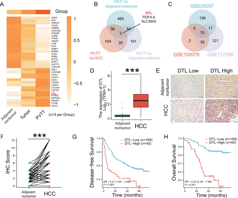
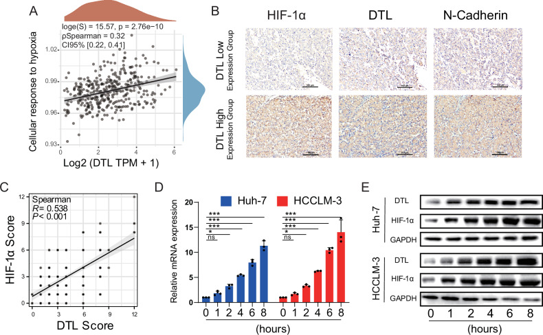
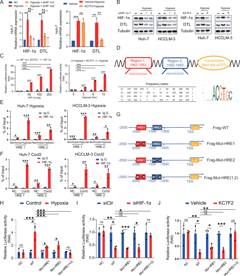
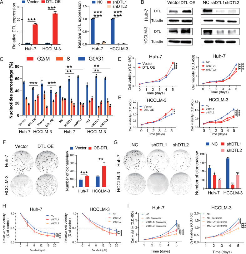
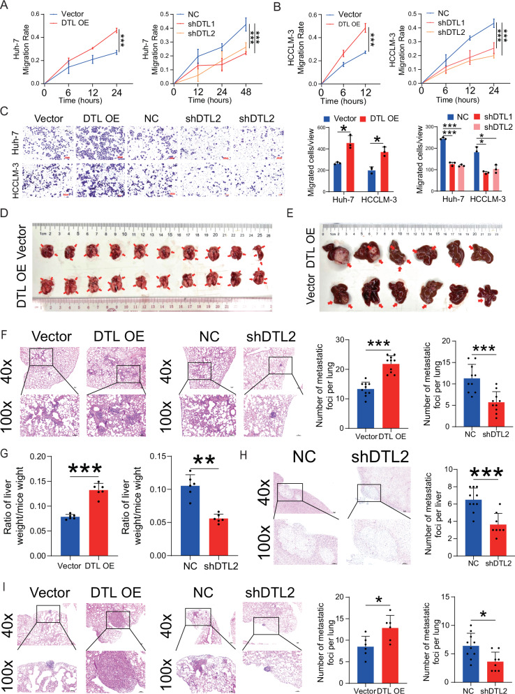
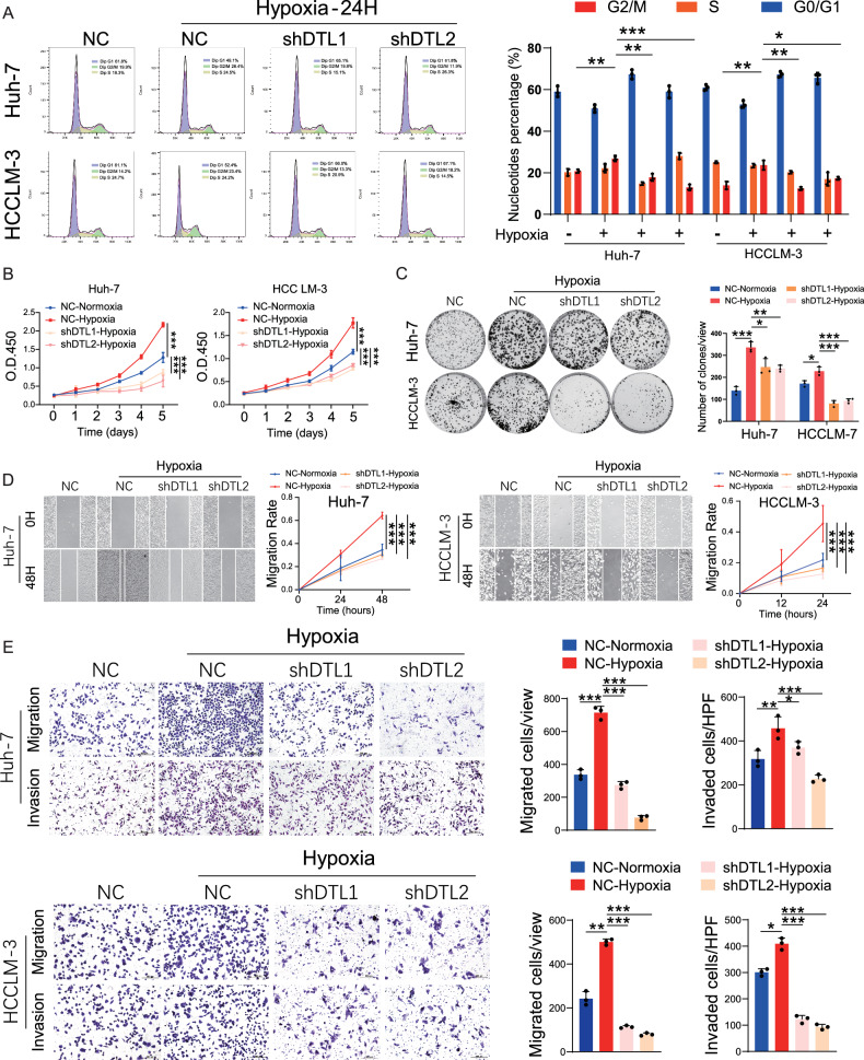
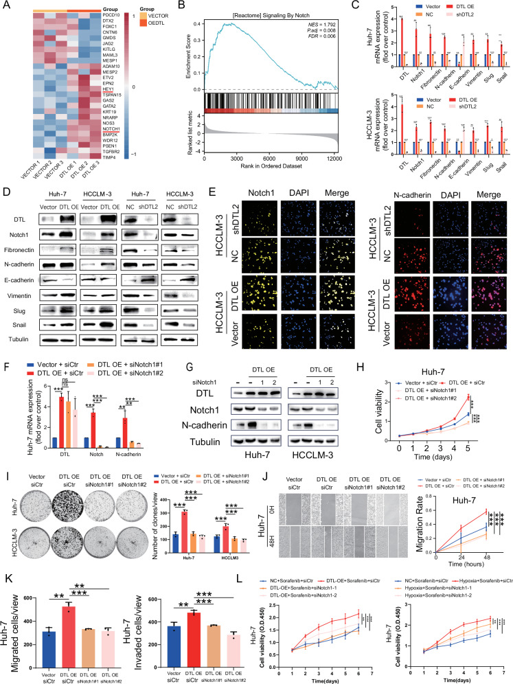
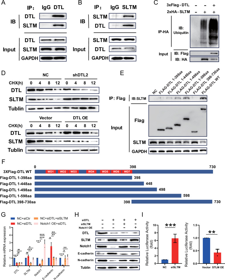
Denticleless E3 ubiquitin protein ligase homolog (DTL), the substrate receptor of the CRL4A complex, plays a central role in genome stability. Even though the oncogenic function of DTL has been investigated in several cancers, its specific role in hepatocellular carcinoma (HCC) still needs further elucidation. Data from a clinical cohort (n = 209), RNA-sequencing, and public database (TCGA and GEO) were analyzed, indicating that DTL is closely related to patient prognosis and could serve as a promising prognostic indicator in HCC. Functionally, DTL promoted the proliferation, metastasis, and sorafenib resistance of HCC in vitro. In the orthotopic tumor transplantation and tail vein injection model, DTL promoted the growth and metastasis of HCC in vivo. Mechanically, we revealed for the first time that DTL was transcriptionally activated by hypoxia-inducible factor 1α (HIF-1α) under hypoxia and functioned as a downstream effector molecule of HIF-1α. DTL promotes the ubiquitination of SAFB-like transcription modulator (SLTM) and subsequently relieves the transcriptional repression of Notch1. These results suggested that DTL may be a potential biomarker and therapeutic target for HCC.


Conflict of interest statement

The authors declare no competing interests.

Figures

Fig. 1
Fig. 1. DTL is overexpressed in human HCCs and correlates with poor prognosis.
A Heatmap depicting mRNA sequencing data from nine samples, which include three pairs of PVTT, tumor, and adjacent non-tumor tissues. The Venn diagrams demonstrate the overlap of differentially expressed genes (DEGs) across these samples (B) and three HCC datasets from GSE databases (C). D DTL transcriptional expression in patients with HCC and adjacent non-tumor from profiling data on TCGA databases. E Representative IHC staining showed the DTL expression level in HCC and adjacent non-tumor tissues (×100). F IHC score analysis of DTL in HCC and adjacent non-tumor samples (n = 209). G, H Disease-free survival and overall survival in DTL-high and DTL-low groups. ***P < 0.001.
Fig. 2
Fig. 2. DTL expression was positively correlated with hypoxia.
A The correlation between DTL and cellular response to the hypoxia pathway was analyzed with Spearman. B Representative IHC staining of HIF-1α, DTL, and N-cadherin in DTL-high and DTL-low HCC tissue. C Correlation analysis between the IHC score of DTL and HIF-1α (n = 209). D The mRNA levels of DTL at different hypoxia time points. E Protein expression of DTL and HIF-1α at different time points. *P < 0.05, ***P < 0.001, ns no significance.
Fig. 3
Fig. 3. HIF-1α activates DTL transcription under hypoxia.
A, B HCC cells were transfected with HIF-1α-siRNA or treated with KC7F2 (40 μM) under hypoxia (1% O2). The mRNA and protein expression of DTL and HIF-1α were determined using qRT-PCR and a western blot assay. C Dual-luciferase reporter assays were performed in HEK-293T cells with HIF-1α overexpression or hypoxia following KC7F2 treatment (40 μM). D Sequences of the predicted HRE in the DTL promoter. E, F Chromatin immunoprecipitation (ChIP) assays in Huh-7 and HCCLM-3 cells to identify the binding of HIF-1α to the DTL promoter under hypoxia or after treatment with CoCl2. G Schematic diagram of the DTL promoter and three mutant constructs. H Dual-luciferase reporter assays for the mutant HRE sequences in HEK-293T cells under hypoxia. I, J Dual-luciferase reporter assays for the mutant HRE sequences in HEK-293T cells following siHIF-1α and KC7F2 (40 μM) under hypoxia. *P < 0.05, **P < 0.01, ***P < 0.001, ns no significance.
Fig. 4
Fig. 4. DTL promotes cell-cycle progression, proliferation, and sorafenib resistance in HCC cells.
A, B DTL overexpression and knockdown stable cell line were established; the DTL mRNA and protein levels were determined by qRT-PCR and western blot. C The effect of DTL overexpression and knockdown on the cell cycle in Huh-7 and HCCLM-3 were analyzed by flow cytometry. D, E The effect of DTL overexpression and knockdown on proliferation in Huh-7 and HCCLM-3 cells was determined by Cell Counting Kit-8 (CCK-8) assays. F, G The effect of DTL overexpression and knockdown on proliferation in Huh-7 and HCCLM-3 cells was determined by clone formation assay. H The relative cell viability of DTL knockdown Huh-7 and HCCLM-3 cells after treatment with different concentrations of sorafenib for 72 h was detected by CCK-8 assays. I CCK-8 assays showed the proliferation of Huh-7 and HCCLM-3 cells response to 3 μM sorafenib with DTL knockdown. *P < 0.05, **P < 0.01, ***P < 0.001, ns no significance.
Fig. 5
Fig. 5. DTL promoted metastasis of HCC cells in vitro and vivo.
A, B The effect of DTL overexpression and knockdown on Huh-7 and HCCLM-3 cells wound healing. C Transwell assays were performed to evaluate the migration ability of DTL overexpression and knockdown HCC cells. D Representative images of lung tissues in tail vein tumor metastasis mouse model from DTL overexpression and vector groups. E Representative images of liver tissues in orthotopic liver transplantation model from DTL overexpression and vector groups. F Representative images of lung metastatic nodules in hematoxylin–eosin (HE) stained sections. The left panel shows the number of lung nodules in DTL overexpression and vector groups from the tail vein tumor metastasis mouse model. G The relative liver weight was calculated as the ratio of wet liver weight to body weight ratio. H Representative images of intrahepatic metastasis nodules in HE‐stained sections. Left panel: the number of intrahepatic metastasis nodules in the DTL knockdown group. I Representative images of lung metastatic nodules in HE‐stained sections. Left panel: the number of lung nodules in DTL overexpression and vector groups from orthotopic liver transplantation model. *P < 0.05, **P < 0.01, ***P < 0.001, ns no significance.
Fig. 6
Fig. 6. The oncogenic role of HIF-1α in HCC cells partly depended on DTL.
A Cell-cycle analysis evaluated the effects of DTL knockdown on cell cycle under hypoxia in HCC cells. B, C Cell Counting Kit-8 (CCK-8) and clone formation assays were performed to evaluate the DTL knockdown on the proliferation of HCC cells under hypoxia. D Representative images and quantification of wound-healing assays for DTL knockdown HCC cells under hypoxia. E Representative images and quantification of transwell migration and invasion assays for DTL knockdown HCC cells under hypoxia. *P < 0.05, **P < 0.01, ***P < 0.001, ns no significance.
Fig. 7
Fig. 7. Notch signaling pathway mediated DTL-induced EMT, proliferation, and metastasis of HCC cells.
A Heatmap of the representative upregulated and downregulated gene after overexpression of DTL in HCCLM-3 cells. B Gene set enrichment analysis (GSEA) of the Notch pathway. C qRT-PCR results of Notch1 and EMT-related genes mRNA expression in DTL overexpressing or knockdown HCC cells. D Western blot results of Notch1 and EMT-related genes protein expression in DTL overexpressing or knockdown HCC cells. E Immunofluorescence results of Notch1 and N-cadherin expression in DTL overexpressing or knockdown HCCLM-3 cells. F, G qRT-PCR and western blot detected the knockdown efficiency of Notch1 by siRNA. H, I The functional role of Notch1 in DTL-induced proliferation of HCC cells was detected by Cell Counting Kit-8 (CCK-8) and clone formation assays. J, K The functional role of Notch1 in DTL-induced metastasis of Huh-7 cells was detected by wound-healing and transwell assays. L CCK-8 assay evaluating Huh-7 cells viability under DTL overexpression or hypoxia with Notch1 knockdown and sorafenib (3 μM) treatment. *P < 0.05, **P < 0.01, ***P < 0.001, ns no significance.
Fig. 8
Fig. 8. DTL directly binds to SLTM and mediates its ubiquitination-dependent degradation.
A, B Co-immunoprecipitation (Co-IP) and western blot (WB) were performed to assess the interaction between exogenous DTL and SLTM in Huh-7 cells. C 2xHA-SLTM was transfected into 293T cells with or without 3xFlag-DTL. The effect of DTL on SLTM ubiquitination was analyzed by IP and WB. D WB analysis of 293T cells with DTL knockdown and those overexpressing DTL, both treated with cycloheximide (CHX, 50 mg/ml) for various time points to evaluate protein degradation. E Co-IP was employed to determine the binding domain of DTL that interacts with SLTM. F Schematic overview of the full-length DTL and its truncated mutants, depicted in plasmid constructs. G, H Quantitative real-time PCR (qRT-PCR) and WB were conducted to assess the expression levels of cleaved Notch1, as well as the Notch1 pathway downstream target genes, in Huh-7 cells. I Luciferase reporter for the Notch1 in 293T cells with SLTM overexpression or knockdown. *P < 0.05, **P < 0.01, ***P < 0.001, ns no significance.

References

    1. Siegel RL, Miller KD, Wagle NS, Jemal A. Cancer statistics, 2023. CA Cancer J Clin. 2023;73:17–48. - DOI - PubMed
    1. Vogel A, Meyer T, Sapisochin G, Salem R, Saborowski A. Hepatocellular carcinoma. Lancet. 2022;400:1345–62. - DOI - PubMed
    1. Yang C, Zhang H, Zhang L, Zhu AX, Bernards R, Qin W, et al. Evolving therapeutic landscape of advanced hepatocellular carcinoma. Nat Rev Gastroenterol Hepatol. 2023;20:203–22. - DOI - PubMed
    1. Zucman-Rossi J, Villanueva A, Nault JC, Llovet JM. Genetic landscape and biomarkers of hepatocellular carcinoma. Gastroenterology. 2015;149:1226–39.e1224. - DOI - PubMed
    1. Corzo CA, Condamine T, Lu L, Cotter MJ, Youn JI, Cheng P, et al. HIF-1α regulates function and differentiation of myeloid-derived suppressor cells in the tumor microenvironment. J Exp Med. 2010;207:2439–53. - DOI - PMC - PubMed

MeSH terms

LinkOut - more resources