. 2024 Oct 9;14(1):172.
doi: 10.1038/s41408-024-01145-0.

DEK regulates B-cell proliferative capacity and is associated with aggressive disease in low-grade B-cell lymphomas

Affiliations

DEK regulates B-cell proliferative capacity and is associated with aggressive disease in low-grade B-cell lymphomas

Melissa A Hopper et al. Blood Cancer J. .

Abstract

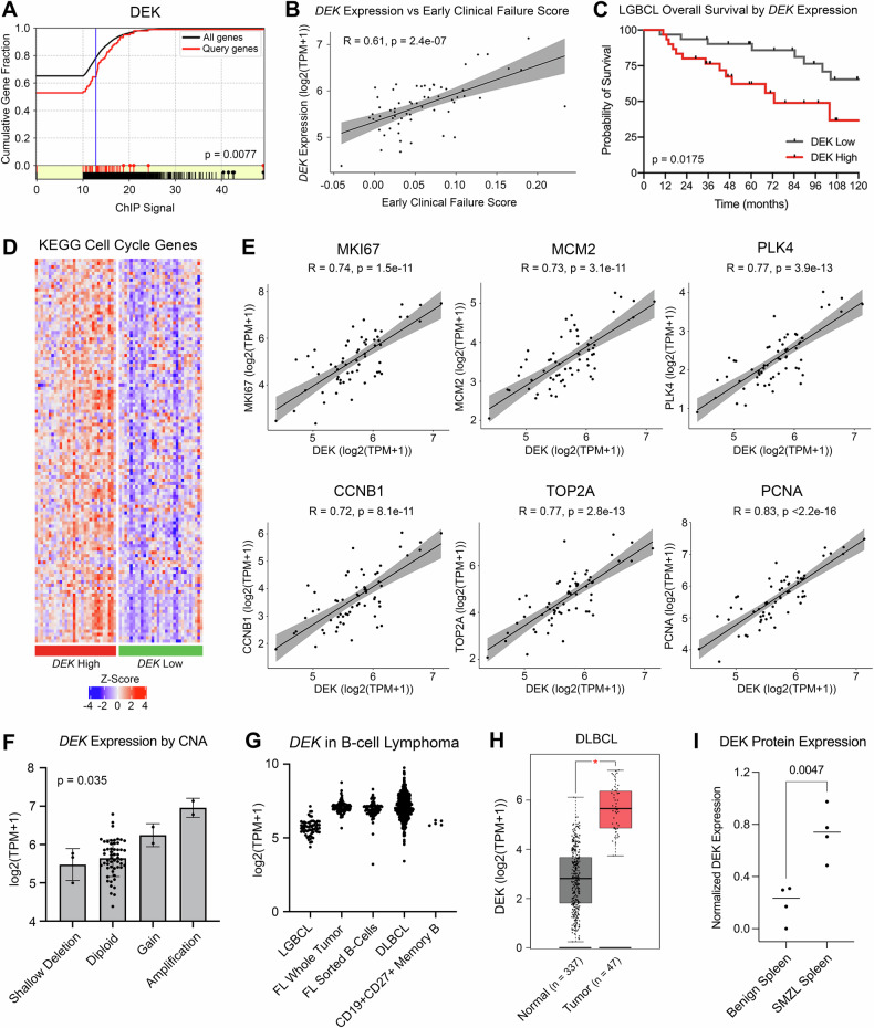
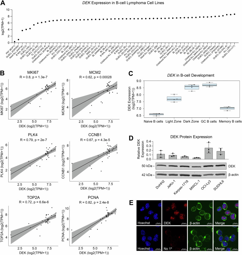
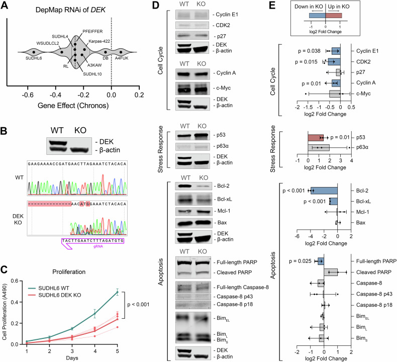
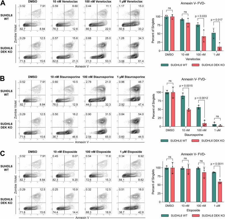
This study sheds light on the pivotal role of the oncoprotein DEK in B-cell lymphoma. We reveal DEK expression correlates with increased tumor proliferation and inferior overall survival in cases diagnosed with low-grade B-cell lymphoma (LGBCL). We also found significant correlation between DEK expression and copy number alterations in LGBCL tumors, highlighting a novel mechanism of LGBCL pathogenesis that warrants additional exploration. To interrogate the mechanistic role of DEK in B-cell lymphoma, we generated a DEK knockout cell line model, which demonstrated DEK depletion caused reduced proliferation and altered expression of key cell cycle and apoptosis-related proteins, including Bcl-2, Bcl-xL, and p53. Notably, DEK depleted cells showed increased sensitivity to apoptosis-inducing agents, including venetoclax and staurosporine, which underscores the therapeutic potential of targeting DEK in B-cell lymphomas. Overall, our study contributes to a better understanding of DEK's role as an oncoprotein in B-cell lymphomas, highlighting its potential as both a promising therapeutic target and a novel biomarker for aggressive LGBCL. Further research elucidating the molecular mechanisms underlying DEK-mediated tumorigenesis could pave the way for improved treatment strategies and better clinical outcomes for patients with B-cell lymphoma.


Conflict of interest statement

AJN has research funding from Bristol Myers Squibb. MAH, ARD, JSW, JPN, MSL, MKM, XW, JEK, VS, MJM, ZY, MDD, TMH, BKL, LMR, TEW, SMA, JRC, and DJ declare no competing financial interests.

Figures

Fig. 1
Fig. 1. DEK is associated with aggressive disease and is impacted by copy number alterations.
A ChIP signal of DEK protein as a regulator of early clinical failure signature genes identified by MAGIC TF analysis. B Correlation of DEK expression from LGBCL tumor RNA-seq data versus early clinical failure signature score. C Kaplan-Meier curves showing overall survival of patients expressing low vs. high levels of DEK. P-values were determined by log-rank test. D Gene expression of KEGG Cell Cycle genes from the Molecular Signatures Database. Heatmap shows z-scored log2(TPM + 1) values from LGBCL tumor RNA-sequencing data, with DEK high cases annotated in red and DEK low cases in green. E Correlation of DEK expression with expression of proliferation markers in RNA-seq data from LGBCL tumors. F DEK expression in LGBCL tumors by copy number status of DEK. G Log2(TPM + 1) expression of DEK across multiple B-cell lymphoma tumor types and healthy donor CD27 + CD19+ sorted memory B-cells. H DLBCL tumor versus normal DEK expression using the DLBC dataset from GEPIA. P-value was calculated in GEPIA using a one-way ANOVA with disease state as the variable for calculating differential expression. I Quantification of DEK expression from western blot analysis comparing benign spleens (n = 4) to SMZL spleens (n = 4). DEK expression levels were normalized to β-actin for each sample.
Fig. 2
Fig. 2. DEK expression is associated with cell lines derived from aggressive B-cell lymphoma proliferative B-cell programs.
A DEK log2(TPM + 1) values in B-cell lymphoma/leukemia cell lines. RNA-seq data from the Broad-Novartis Cell Line Encyclopedia was acquired through the DepMap Portal. B Correlation of DEK expression with expression of proliferation markers in cell line RNA-seq data from Fig. 2A. C DEK log2(TPM + 1) values across B-cell development stages. D Western blot of DEK protein expression across B-cell lymphoma cell lines. Bar plot represents DEK expression normalized to β-actin. Representative blot of n = 3 biological replicates, with quantification of all blots shown in the bar plot. Blot has been cropped to display indicated proteins based on molecular weight. E DEK localization in B-cell lymphoma cell line SUDHL6 by confocal microscopy.
Fig. 3
Fig. 3. DEK depletion inhibits proliferation and is accompanied by reduced expression of cell cycle genes, reduced Bcl-2 and Bcl-xL expression, and increased p53 expression.
A Chronos Gene Effect from RNAi of DEK in B-cell lymphoma cell lines. Gene effect represents the impact of DEK RNAi on cellular fitness, with negative values representing a negative impact to cellular fitness. B Validation of DEK knockout by western blot and Sanger sequencing. C In vitro five-day proliferation assay of synchronized SUDHL6 WT versus DEK KO cells. Proliferation was measured by MTT assay with n = 3 biological replicates. Assays were run with technical triplicates. D Western blot analysis of cell cycle, apoptosis, and stress response proteins in SUDHL6 WT and DEK KO cells. Cell lines were harvested at ≥ 95% viability. Blots have been cropped to display indicated proteins based on molecular weight. The corresponding DEK and β-actin controls were processed in parallel and are displayed below their corresponding samples. E Quantifications of protein expression in DEK KO versus WT cells of n = 3 biological replicates. All quantifications were scaled to β-actin levels and fold-changes of DEK KO versus WT were used as input for statistical analysis. Plots depict the mean and SD of log2 fold change values, where negative values represent reduced expression in DEK KO. Blue bars indicate a statistically significant decrease of expression in DEK KO cells, red bars indicate a statistically significant increase in DEK KO cells.
Fig. 4
Fig. 4. DEK knockout cells are more susceptible to apoptosis and demonstrate increased sensitivity to venetoclax treatment.
Annexin V and Zombie Violet staining of SUDHL6 WT versus DEK KO cells treated with the indicated concentrations of (A) venetoclax, (B) staurosporine, and (C) etoposide. Cells were treated for 24 h at 37 °C, stained with Annexin V-FITC and Zombie Violet FVD, and gated on singlets. Bar charts show the mean and SD of frequencies of viable cells (Annexin V-FVD-) from n = 3 biological replicates.

References

    1. Alaggio R, Amador C, Anagnostopoulos I, Attygalle AD, Araujo IBDO, Berti E, et al. The 5th edition of the World Health Organization Classification of Haematolymphoid Tumours: Lymphoid Neoplasms. Leukemia. 2022;36:1720–48. - PMC - PubMed
    1. Smyth E, Cheah CY, Seymour JF. Management of indolent B-cell Lymphomas: A review of approved and emerging targeted therapies. Cancer Treat Rev. 2023;113:102510. - PubMed
    1. Tracy SI, Larson MC, Feldman AL, Maurer MJ, Novak AJ, Slager SL, et al. The utility of prognostic indices, early events, and histological subtypes on predicting outcomes in non-follicular indolent B-cell lymphomas. Am J Hematol. 2019;94:658–66. - PMC - PubMed
    1. Cerhan JR, Link BK, Habermann TM, Maurer MJ, Feldman AL, Syrbu SI, et al. Cohort Profile: The Lymphoma Specialized Program of Research Excellence (SPORE) Molecular Epidemiology Resource (MER) Cohort Study. Int J Epidemiol. 2017;46:1753–1754i. - PMC - PubMed
    1. Hopper MA, Wenzl K, Hartert KT, Krull JE, Dropik AR, Novak JP, et al. Molecular classification and identification of an aggressive signature in low-grade B-cell lymphomas. Hematological Oncol. 2023;41:644–54. - PMC - PubMed

Publication types

MeSH terms

Substances

LinkOut - more resources