. 2024 Oct 10;14(1):23653.
doi: 10.1038/s41598-024-71770-z.

Antifibrotic effect of disulfiram on bleomycin-induced lung fibrosis in mice and its impact on macrophage infiltration

Affiliations

Antifibrotic effect of disulfiram on bleomycin-induced lung fibrosis in mice and its impact on macrophage infiltration

Yugo Okabe et al. Sci Rep. .

Abstract

The accumulation of monocyte-derived macrophages in the lung tissue during inflammation is important for the pathogenesis of fibrotic lung disease. Deficiencies in chemokine receptors CCR2 and CCR5 and their ligands, which mediate monocyte/macrophage migration, ameliorate bleomycin (BLM)-induced lung fibrosis. Disulfiram (DSF), which is used to treat alcoholism because of its aldehyde dehydrogenase (ALDH)-inhibiting effect, inhibits monocyte/macrophage migration by inhibiting FROUNT, an intracellular regulator of CCR2/CCR5 signalling. Here, we investigated the antifibrotic effect of oral DSF administration in a mouse model of BLM-induced lung fibrosis, focusing on macrophage response and fibrosis progression. The direct inhibitory activity of DSF on monocyte migration was measured using the Boyden chamber assay and compared with that of DSF-related inhibitors with different FROUNT-inhibition activities. Quantitative PCR was used to determine the expression of fibrosis-promoting genes in the lung tissue. DSF significantly suppressed macrophage infiltration into lung tissues and attenuated BLM-induced lung fibrosis. DSF and its metabolites, diethyldithiocarbamate (DDC) and copper diethyldithiocarbamate (Cu(DDC)2), inhibited monocyte migration toward the culture supernatant of primary mouse lung cells mainly comprising CCL2, whereas cyanamide, another ALDH inhibitor, did not. DSF, with higher inhibitory activity against FROUNT than DDC and Cu(DDC)2, inhibited monocyte migration most strongly. In BLM-induced fibrotic lung tissues, profibrotic factors were highly expressed but were reduced by DSF treatment. These results suggest DSF inhibits macrophage infiltration, which might be attributed to its inhibitory effect on FROUNT, and attenuates BLM-induced lung fibrosis. In addition, multiplex immunofluorescence imaging revealed reduced infiltration of S100A4+ macrophages into the lungs in DSF-treated mice and high expression of FROUNT in S100A4+ macrophages in idiopathic pulmonary fibrosis (IPF). These findings underscore the potential of macrophage-targeted therapy with DSF as a promising drug repositioning approach for treating fibrotic lung diseases, including IPF.


Conflict of interest statement

The authors declare no competing interests.

Figures

Fig. 1
Fig. 1
Effect of DSF on BLM-induced lung fibrosis in mice. (A) Time course of percentage change in body weight following BLM administration. (B) SP-D concentration in the blood of mice on days 0, 14, and 28. (CH) Representative cross-sectional micro-CT images of mouse lungs on days 14 (C, D, E) and 28 (F, G, H) in the untreated (C, F), DSF non-treated (D, G), and DSF-treated (E, H) groups. (I) Percentage of the abnormally hyperabsorbed area (between − 400 and − 200 Hounsfield units) in the whole lung field on days 0, 14, and 28. n = 5 per group. *P < 0.05, **P < 0.01. Sidak’s multiple comparison test for (A) and Tukey’s test for (B) and (I) were used to compare DSF non-treated and DSF-treated groups.
Fig. 2
Fig. 2
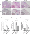
Histological analysis and hydroxyproline content of BLM-induced lung fibrosis in mice. (AF) Haematoxylin and eosin staining (AC) and Elastica Masson–Goldner (EMG) staining (DF) of lung tissues on day 28 in untreated (A, D), DSF non-treated (B, E), and DSF-treated (C, F) mice. Higher magnification images in the lower left correspond to the areas indicated by blue squares. Scale bars = 500 μm. (G) Number of infiltrating cells in the lung tissue on day 28 counted in HE images (AC). (H) Ashcroft score of lung tissues on day 28 evaluated in EMG images (DF). (I) Hydroxyproline content in the lung tissue on day 28. n = 5 per group. *P < 0.05, **P < 0.01 (Tukey’s test). ns, not significant.
Fig. 3
Fig. 3
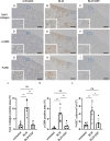
Immunohistochemical analysis of BLM-induced lung fibrosis in mice. (AF) Representative immunostaining images for type I collagen (AC), α-SMA (DF), and F4/80 (GI) in the lung tissue of untreated (A, D, G), DSF non-treated (B, E, H) and DSF-treated (C, F, I) mice on day 28. Higher-magnification images in the lower left correspond to the areas indicated by blue squares. Scale bars = 500 μm. (J, K) Percentage of type I collagen positive and α-SMA positive area in lung tissue on day 28. (L) Number of F4/80+ cells in the lung tissue on day 28 counted in immunostaining images (GI). n = 5 per group. *P < 0.05, **P < 0.01 (Tukey’s test). ns, not significant.
Fig. 4
Fig. 4
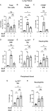
Flow cytometric analysis of lung tissues and peripheral blood of BLM-induced lung fibrosis in mice. (A) Total number of viable cells in the lung tissue of untreated, DSF non-treated, and DSF-treated mice on day 28. (BF) Percentages of total monocytes/macrophages, CD86+ M1 macrophages, CD206+ M2 macrophages, Ly-6Chi monocytes, and neutrophils among total viable cells in the lung tissue of untreated, DSF non-treated, and DSF-treated mice on day 28. (G, H) Percentages of monocytes and neutrophils in total viable cells in the peripheral blood of untreated, DSF non-treated, and DSF-treated mice on day 28. n = 5 per group. *P < 0.05, **P < 0.01 (Tukey’s test). ns, not significant.
Fig. 5
Fig. 5
Effect of DSF on monocyte migration in vitro. (A) Schematic diagram of the Boyden chamber-based monocyte chemotaxis assay toward lung cell culture supernatant (LCCS). Monocytes in input cells and migrated cells were quantified by flow cytometry, and the migration efficiency (number of monocytes in migrated cells/number of monocytes in input cells [%]) was calculated. (B) Migration efficiency of monocytes after CCR2 inhibitor (BMS CCR2 22) pretreatment. n = 3 or 4 per group. (C) Concentrations of 13 chemokines (CXCL1, CXCL5, CXCL9, CXCL10, CXCL13, CCL2, CCL3, CCL4, CCL5, CCL11, CCL17, CCL20, and CCL22) as migration factors in the supernatant of lung tissue-derived cells of C57BL/6J mice after 72 h of culture. (D) Migration efficiency of monocytes pretreated with DSF, DDC, Cu(DDC)2, and cyanamide (ALDH inhibitor). n = 3 per group. (E) Percentage of viable bone marrow cells pretreated with DSF, DDC, Cu(DDC)2, and cyanamide (ALDH inhibitor). The percentage of viable cells not treated with inhibitors was used as the reference (100%). n = 3 per group. *P < 0.05, **P < 0.01 (Tukey’s test).
Fig. 6
Fig. 6
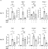
Effect of DSF on profibrotic gene expression in the lung tissues of BLM-induced lung fibrosis in mice. (A, B) Relative mRNA expression of Ccl2, Spp1, Col1a1, and Timp-1 in the lung tissue of untreated, DSF non-treated and DSF-treated mice on day 14 (A) and day 28 (B). n = 4 or 5 per group. *P < 0.05, **P < 0.01 (Tukey’s test). ns, not significant.
Fig. 7
Fig. 7
Effect of DSF on S100A4+ macrophages in the lung tissues of BLM-induced lung fibrosis in mice. (AC) Representative immunostaining images for S100A4 in the lung tissue of untreated (A), DSF non-treated (B) and DSF-treated (C) mice on day 28. Higher-magnification images in the lower left correspond to the areas indicated by dashed squares. Scale bars = 200 μm. (D) Representative bright-field and double-fluorescence staining images in the lung tissues of BLM-treated mice on day 28. DAPI (4′,6-diamidino-2-phenylindole) is shown in blue, CD68 in green, and S100A4 in red. The bright-field image was generated from fluorescent images using inForm software. Higher-magnification images correspond to the area indicated with dashed squares. Arrowheads indicate S100A4+CD68+ macrophages. Scale bars = 25 μm. (E) Number of S100A4+ cells in the lung tissue on day 28 counted in immunostaining images (AC). n = 5 per group. *P < 0.05, **P < 0.01 (Tukey’s test). ns, not significant.
Fig. 8
Fig. 8
Analysis of multi-fluorescent staining of lung tissues of IPF. (A, B) Representative immunofluorescent staining images in normal lung (A) and in fibrotic areas of the lungs of IPF patients (B). DAPI is shown in pink, α-SMA in brown, CD34 in light blue, CD68 in green, S100A4 in red, and FROUNT in blue. Higher-magnification images on the right correspond to the area indicated with dashed squares. White arrows indicate CD68+ macrophages in normal lung. White arrowheads indicate S100A4+CD68+ macrophages and yellow arrowheads indicate S100A4-CD68+ macrophages in fibrotic areas of the lungs of IPF patients. Scale bars = 25 μm. (C) S100A4 signals of each CD68+ macrophage in normal lung tissues and in fibrotic areas of the lungs of IPF patients. (D) FROUNT signals in S100A4-CD68+ macrophages and S100A4+CD68+ macrophages in fibrotic areas of the lungs of IPF patients. n = 5 or 6 per group. **P < 0.01 (unpaired t-test).

References

    1. Kolahian, S., Fernandez, I. E., Eickelberg, O. & Hartl, D. Immune mechanisms in pulmonary fibrosis. Am. J. Respir. Cell Mol. Biol.55, 309–322. 10.1165/rcmb.2016-0121TR (2016). - PubMed
    1. Ishida, Y. et al. Essential roles of the CC chemokine ligand 3-CC chemokine receptor 5 axis in bleomycin-induced pulmonary fibrosis through regulation of macrophage and fibrocyte infiltration. Am. J. Pathol.170, 843–854. 10.2353/ajpath.2007.051213 (2007). - PMC - PubMed
    1. Okuma, T. et al. C-C chemokine receptor 2 (CCR2) deficiency improves bleomycin-induced pulmonary fibrosis by attenuation of both macrophage infiltration and production of macrophage-derived matrix metalloproteinases. J. Pathol.204, 594–604. 10.1002/path.1667 (2004). - PubMed
    1. Baran, C. P. et al. Important roles for macrophage colony-stimulating factor, CC chemokine ligand 2, and mononuclear phagocytes in the pathogenesis of pulmonary fibrosis. Am. J. Respir. Crit. Care Med.176, 78–89. 10.1164/rccm.200609-1279OC (2007). - PMC - PubMed
    1. McCubbrey, A. L. et al. Deletion of c-FLIP from CD11b hi macrophages prevents development of bleomycin-induced lung fibrosis. Am. J. Respir. Cell Mol. Biol.58, 66–78. 10.1165/rcmb.2017-0154OC (2018). - PMC - PubMed

MeSH terms