Review
. 2024 Dec;24(12):887-905.
doi: 10.1038/s41568-024-00750-2. Epub 2024 Oct 9.

Steering research on mRNA splicing in cancer towards clinical translation

Affiliations
Review

Steering research on mRNA splicing in cancer towards clinical translation

Olga Anczukow et al. Nat Rev Cancer. 2024 Dec.

Abstract

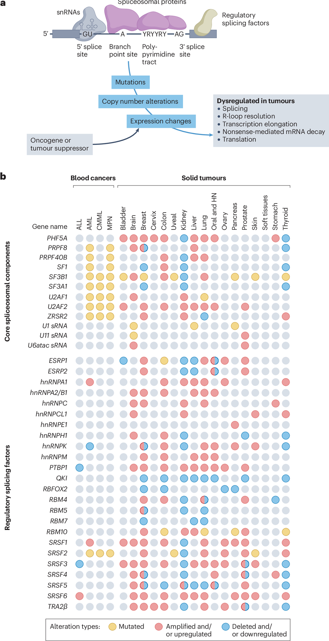
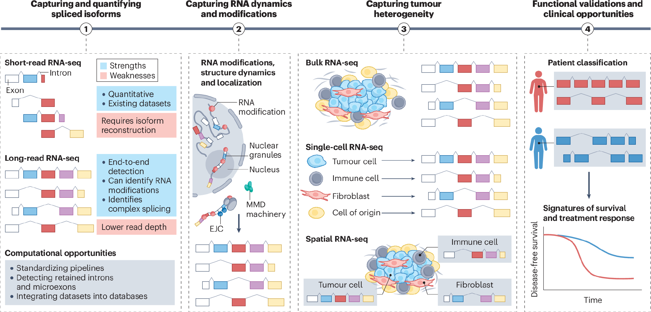
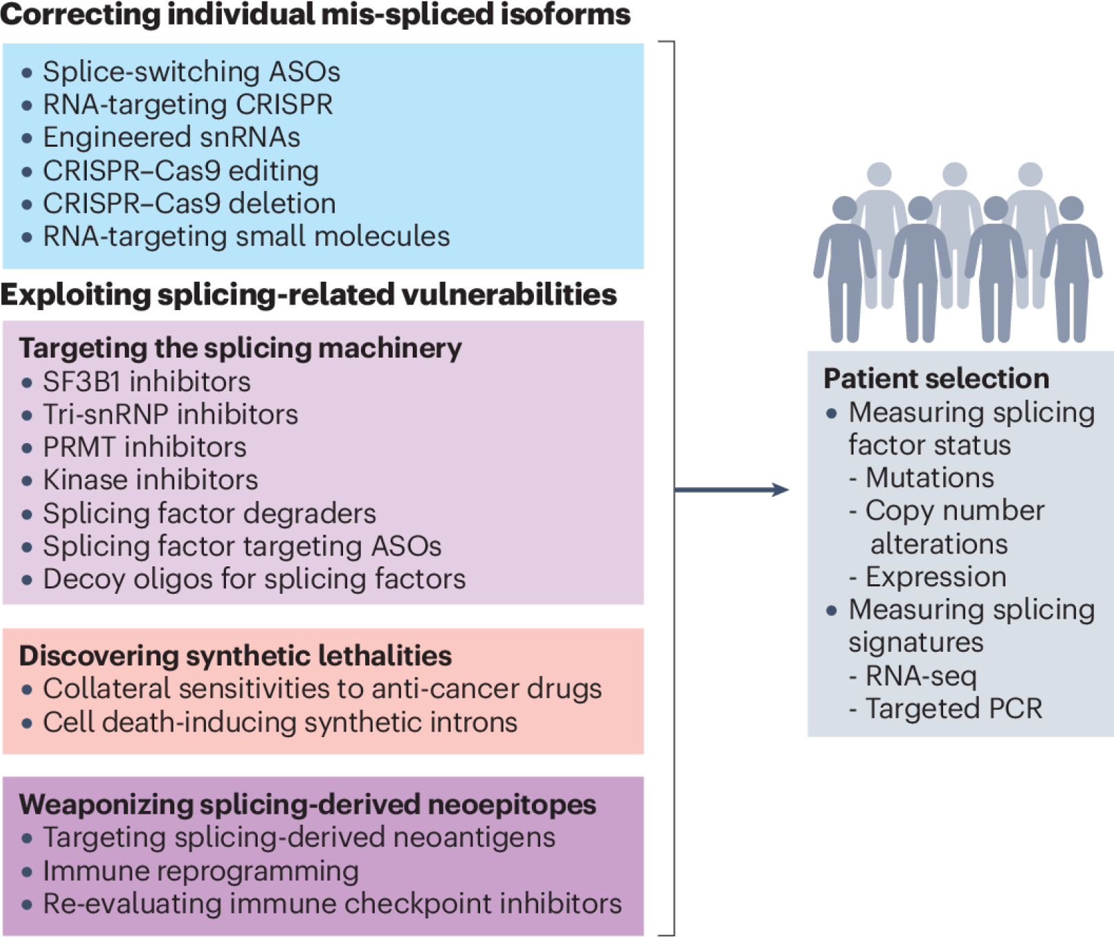
Splicing factors are affected by recurrent somatic mutations and copy number variations in several types of haematologic and solid malignancies, which is often seen as prima facie evidence that splicing aberrations can drive cancer initiation and progression. However, numerous spliceosome components also 'moonlight' in DNA repair and other cellular processes, making their precise role in cancer difficult to pinpoint. Still, few would deny that dysregulated mRNA splicing is a pervasive feature of most cancers. Correctly interpreting these molecular fingerprints can reveal novel tumour vulnerabilities and untapped therapeutic opportunities. Yet multiple technological challenges, lingering misconceptions, and outstanding questions hinder clinical translation. To start with, the general landscape of splicing aberrations in cancer is not well defined, due to limitations of short-read RNA sequencing not adept at resolving complete mRNA isoforms, as well as the shallow read depth inherent in long-read RNA-sequencing, especially at single-cell level. Although individual cancer-associated isoforms are known to contribute to cancer progression, widespread splicing alterations could be an equally important and, perhaps, more readily actionable feature of human cancers. This is to say that in addition to 'repairing' mis-spliced transcripts, possible therapeutic avenues include exacerbating splicing aberration with small-molecule spliceosome inhibitors, targeting recurrent splicing aberrations with synthetic lethal approaches, and training the immune system to recognize splicing-derived neoantigens.


Conflict of interest statement

Competing interests: J.V. is a member of the Advisory Boards of Remix Therapeutics, Stoke Therapeutics and IntronX. K.M.W. is an adviser to and holds equity in Ribometrix, ForagR Medicines and A-Form Solutions. O.A. is a member of the Advisory Boards of Caeruleus Genomics.

Figures

Figure 1.
Figure 1.. Splicing factor alterations in human tumors.
a, Core spliceosomal proteins (purple) and small nuclear RNAs (snRNAs) (grey) are critical components of constitutive RNA splicing, whereas regulatory splicing factors (tan) have a role in alternative splicing. These are depicted assembled on a pre-mRNA molecule composed of exons (grey rectangles) and introns (black lines). The 5′ and 3′ splice sites are depicted along with highly conserved dinucleotides GU and AG, which define intron boundaries, the adenosine residue serving as the branch point site, and the polypyrimidine tract. Tumours exhibit splicing factor mutations, copy number alterations, or expression changes. In some tumours, these splicing factor expression changes are induced directly or indirectly by oncogenes or tumour suppressors. Splicing factor alterations lead to dysregulation of alternative splicing but also of other processes listed on the right. b, Genomic events that affect expression and function of selected splicing factors in blood (left) and solid (right) cancers. They affect both select core (top) and regulatory (bottom) spliceosome components. The data presented have been curated from the literature and represent key splicing factors that are frequently altered in primary or metastatic human tumours compared with normal tissues (see references in Supplementary Table 1). Splicing factors can be mutated (single nucleotide changes), upregulated with or without gene amplification, or downregulated with or without gene deletion. A limitation of these data is that given their non-focal nature, amplification and shallow deletions are more difficult to functionally interpret than deep deletions. Note that for some splicing factors, individual studies have reported them to be either upregulated or downregulated in the same tumour type. These discrepancies can often be explained by differences in tumour subtypes, tumour stages, and even spatial localization within the tumour tissues (for example, carcinoma in situ versus the invasive tumour front). ALL, acute lymphoblastic leukaemia; AML, acute myeloid leukaemia; CMML, chronic myelomonocytic leukaemia; HN, head and neck cancers; MPN, myeloproliferative neoplasms.
Figure 2.
Figure 2.. Four key steps towards clinical translation of mRNA splicing research.
The first step towards translating mRNA splicing research is to capture and quantify isoforms. The strengths and weaknesses are listed for current RNA-sequencing (RNA-seq) approaches, along with computational challenges that remain be addressed. Second, understanding the role of RNA processing in cancer requires profiling of complex repertoires of RNA isoforms, their structure and folding, any chemical modifications (including m7G, N6-methyladenosine (m6A) and m5C ψ), A-I editing (represented by red bubbles), the cellular localization (to specific nuclear comportments or phase-separated granules), the stability and turnover (including cytoplasmic degradation by the nonsense mediated mRNA decay (NMD) pathway of transcripts containing an exon junction complex (EJC) downstream of their stop codon), and the RNA dynamics. All of these factors can impact splicing, stability, nuclear export and translation. Third, capturing spliced isoform heterogeneity and their spatial localization will enable isoforms expressed in tumour cells to be distinguished from those originating in the tumour microenvironment. This is critical for splicing-directed therapies that are currently being developed, often with the assumption that all cells within the tumour express the same isoform. Lastly, identifying spliced isoforms in tumours offers multiple clinical opportunities. For example, classifying patients based on their isoform profile might reveal novel tumour subtypes, some of which might be associated with distinct survival patterns, prognosis, and drug responses.
Figure 3.
Figure 3.. Strategies that target mRNA splicing in cancer.
Transcript-centric therapeutic strategies (blue box) target individual mis-spliced mRNAs that are expressed in tumour cells, primarily to alter their splicing patterns. These strategies yield distinct protein isoforms, which either gain tumour-suppressive properties or lose oncogenic ones. Alternatively, they yield non-productive transcripts that are degraded by nonsense-mediated mRNA decay (NMD). Splice-switching can be achieved, for example, by using antisense oligonucleotides (ASOs) that block splicing regulatory elements. Similarly, exogenous small nuclear RNAs (snRNAs) can be engineered to match specific splice sites and in doing so increase inclusion ‘weak’ exons. For genome editing, Cas9 proteins are often paired with single guide RNAs (sgRNAs) to delete a specific exon in a given transcript or to base-edit a specific splice site to restore or to compromise its utilization, leading to exon inclusion or skipping, respectively. Finally, recent studies reported the discovery of small molecules that can recognize a specific RNA sequence or structure and modulate splicing of a specific exon. Spliceosome-centric strategies (light purple box), focus on proteins that controls splicing of multiple downstream transcripts, instead of targeting specific RNA isoforms. These approaches include small-molecule inhibitors that block the activity of the core spliceosomal protein splicing factor 3b subunit 1 (SF3B1), prevent the recruitment of the U4/U5/U6 tri-snRNP, or block the activity of protein kinases or protein arginine methyltransferases (PRMTs) which regulate the activity of splicing factors at the post-translational level. Alternatively, the protein levels of a specific splicing factor can be directly targeted using dedicated protein degraders or splice-switching ASOs that, by modulating poison exon skipping or inclusion, can increase or decrease splicing factor protein levels. Finally, an ASO with sequence complementarity to a splicing factor binding motif can be used as decoy to sequester the splicing factor away from its cognate mRNA targets. In parallel, numerous approaches can exploit synthetic lethalities (pink box), as specific splicing alterations often create new vulnerabilities, including sensitizing cancer cells to conventional anti-cancer drugs. Alternatively, a set of specific introns were found to be spliced exclusively in cancer cells with SF3B1 mutations, opening the door to the use of synthetic intron sequences to enable SF3B1 mutation-dependent expression of exogenous cell death-inducing genes. The last class of splicing-based therapies leverages splicing-derived neo-epitopes produced by cancer cells or even creates new ones (dark purple box). These strategies often involve chimeric antigen receptor (CAR) T cells that target abnormally expressed surface protein isoforms or induce immune reprogramming by selectively promoting spliced isoforms associated with enhanced cytotoxicity, cytokine secretion and the generation of memory T cells. The success of these strategies in the clinic will require appropriate patient selection, which should include more systematic molecular measurements of both the splicing factor status and RNA isoform signatures (grey box). This will help to identify patients who would benefit from therapies targeting specific mRNA isoforms versus those that would respond to more global approaches exploiting splicing-related vulnerabilities. RNA-seq, RNA sequencing; snRNP, small nuclear ribonucleoprotein.

References

    1. Berget SM, Moore C & Sharp PA Spliced segments at the 5’ terminus of adenovirus 2 late mRNA. Proc Natl Acad Sci U S A 74, 3171–3175 (1977). - PMC - PubMed
    1. Chow LT, Gelinas RE, Broker TR & Roberts RJ An amazing sequence arrangement at the 5’ ends of adenovirus 2 messenger RNA. Cell 12, 1–8 (1977). - PubMed
    1. Pan Q, Shai O, Lee LJ, Frey BJ & Blencowe BJ Deep surveying of alternative splicing complexity in the human transcriptome by high-throughput sequencing. Nat Genet 40, 1413–1415 (2008). - PubMed
    1. Wang ET et al. Alternative isoform regulation in human tissue transcriptomes. Nature 456, 470–476 (2008). - PMC - PubMed
    1. Kjer-Hansen P & Weatheritt RJ The function of alternative splicing in the proteome: rewiring protein interactomes to put old functions into new contexts. Nat Struct Mol Biol 30, 1844–1856 (2023). - PubMed