. 2024 Oct 9;14(1):23623.
doi: 10.1038/s41598-024-74757-y.

Rejuvenation of human mesenchymal stem cells using a nonintegrative and conditionally removable Sendai virus vector

Affiliations

Rejuvenation of human mesenchymal stem cells using a nonintegrative and conditionally removable Sendai virus vector

Mitsuo Oshimura et al. Sci Rep. .

Abstract

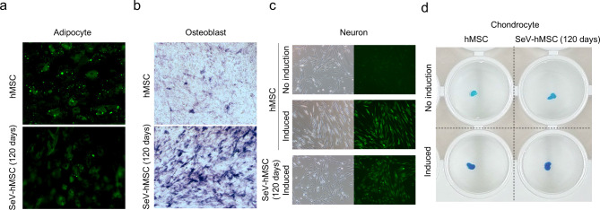
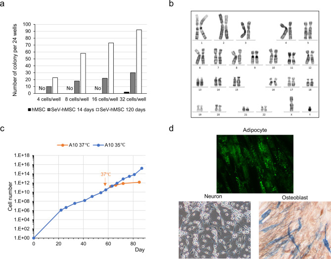
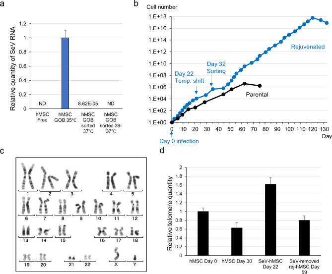
Human mesenchymal stem cells (hMSCs) with extended lifespan and differentiation potential that can recapitulate in vivo characteristics could significantly contribute to basic research, drug development, and cell therapy. Specifically, they could ensure a stable supply of specific cellular resources, and possibly extracellular vesicles. Here, we established a technology for extending the lifespan while maintaining differentiation potential, termed "rejuvenation," of hMSCs (rej-hMSCs) using nonintegrative and conditionally removable temperature-sensitive Sendai virus (SeV) vectors. Various immortalizing factors (i.e., Bmi-1, hTERT, SV40T, and/or HPV E6/E7) were first introduced by the SeV vector into the cells. A combination of three SeVs with Bmi-1, hTERT, or SV40T conferred markedly improved cell proliferation and cloning ability while maintaining differentiation potential and a normal karyotype. An extended lifespan was also demonstrated in other cell types. The rejuvenation of long-passaged or aged hMSCs was also confirmed. SeV vectors were rapidly removed as a function of cell doubling by increasing the temperature from 35 °C to 37 °C or higher, while proliferative ability was maintained. Following FACS sorting, the complete removal of SeV vectors was confirmed by qPCR analyses. Therefore, our cell rejuvenation technology could contribute to research and clinical applications by enabling the supply of modified cells without damaging host chromosomes.

Keywords: Extended lifespan; Human mesenchymal stem cell; Regenerative medicine; Rejuvenation; Sendai virus vector.


Conflict of interest statement

M.O. is a CEO, employee, and shareholder of Trans Chromosomics, Inc., and I.K., T.E., and K.H. are employees of Trans Chromosomics, Inc. The remaining authors declare that they have no conflicts of interest.

Figures

Fig. 1
Fig. 1
Biological effects following transfection of SeV(Bmi-1/hTERT/SV40T) and temperature change. a Proliferation curves of Bmi-1/hTERT/SV40T-transfected cells: 35 °C culture of hMSC parental cell line, 35 °C culture of SeV-infected hMSCs (SeV-hMSCs), and SeV-hMSCs subjected to temperature change from 35 °C to 37 °C. The green arrow indicates the point at which the temperature was changed from 35 °C to 37 °C. b Transmitted light (BF) and fluorescence (OFP, GFP) images of cells transfected with three factors (Bmi-1/hTERT/SV40T) are shown: culture maintained at 35 °C and after temperature change from 35 °C to 37 °C. c Transmitted light images of SeV(Bmi-1/hTERT/SV40T)-hMSCs in culture maintained at 35 °C and after temperature change from 35 °C to 37 °C. d Changes in telomere length after transfection with Bmi-1/hTERT/SV40T, and with and without removing the immortalization factors by temperature change. *p < 0.001. e Representative karyotype image of SeV(Bmi-1/hTERT/SV40T)-infected cells after 90 days of passaging (normal karyotype).
Fig. 2
Fig. 2
Differentiation potential of SeV(Bmi-1/hTERT/SV40T)-infected cells. Images of a adipocytes, b osteoblasts, c neurons, and d chondrocytes differentiated from parental and SeV-hMSCs.
Fig. 3
Fig. 3
Cloning of cells. a Comparison of the colony-forming ability in parental hMSCs and SeV-hMSCs after 14 and 120 days of infection. b Representative karyotype image of cloned SeV-hMSCs. c Proliferation curve of representative cloned MSCs. d Representative images of differentiated cloned SeV-hMSCs (adipocyte differentiation, neuron differentiation, osteoblast culture).
Fig. 4
Fig. 4
Growth and morphology of a single clone isolated at different cell ages. a Red arrows indicate cryopreservation points of hMSCs for single-cell cloning test (hMSCs at early, mid-, late, and cell growth-arrested stages on days 9, 24, 49, and 72, respectively). Black arrows indicate points of SeV vector infection (hMSCs at early, mid-, late, and cell growth-arrested stages on days 21, 36, 61, and 90, respectively). b Photographs of SeV vector-infected cells (early-, mid-, and late-stage hMSCs) and SeV vector-uninfected hMSCs (control) before single-cell cloning. c Images of SeV infection test results on mitotic cells (growth arrested-stage hMSCs, day 90): cells immediately before SeV infection, SeV vector-infected cells (2 weeks after infection date), and SeV vector-uninfected cells cultured for 2 weeks (control).
Fig. 5
Fig. 5
Analyses of SeV-hMSCs following removal of the SeV vector. a RT-qPCR results for SeV vector in parental hMSCs (hMSC Free), SeV-hMSCs (hMSC GOB 35 °C), and SeV-hMSCs cultured at 37 °C after sorting from 37 °C (hMSC GOB sorted 37 °C) or 39/37°C culture (hMSC GOB sorted 39 –37 °C). b Growth curve and accumulative cell number of SeV-hMSCs and SeV-removed hMSCs. The temperature was shifted on day 22 and cell sorting of fluorescence-negative cells was performed on day 32. c Representative karyotype image of SeV-removed hMSCs. d Telomere length analysis of hMSCs at each stage.

Similar articles

Cited by

References

    1. Lim, W. A. The emerging era of cell engineering: harnessing the modularity of cells to program complex biological function. Science. 378, 848–852 (2022). - PubMed
    1. Khademi-Shirvan, M., Ghorbaninejad, M., Hosseini, S. & Eslaminejad, M. B. The importance of stem cell senescence in regenerative medicine. Adv. Exp. Med. Biol. 1288, 87–102 (2020). - PubMed
    1. Li, Y. et al. Senescence of mesenchymal stem cells (review). Int. J. Mol. Med. 39, 775–782 (2017). - PubMed
    1. Dunn, C. M., Kameishi, S., Grainger, D. W. & Okano, T. Strategies to address mesenchymal stem/stromal cell heterogeneity in immunomodulatory profiles to improve cell-based therapies. Acta Biomater. 133, 114–125 (2021). - PubMed
    1. Tátrai, P. et al. Combined introduction of Bmi-1 and hTERT immortalizes human adipose tissue-derived stromal cells with low risk of transformation. Biochem. Biophys. Res. Commun. 422, 28–35 (2012). - PubMed

LinkOut - more resources