. 2024 Oct 9;7(1):1290.
doi: 10.1038/s42003-024-06997-7.

HES1 potentiates high salt stress response as an enhancer of NFAT5-DNA binding

Affiliations

HES1 potentiates high salt stress response as an enhancer of NFAT5-DNA binding

Hiroki Ryuno et al. Commun Biol. .

Abstract

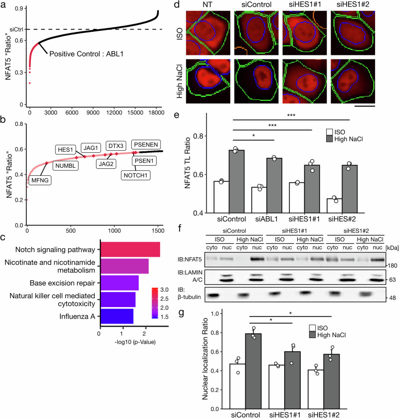
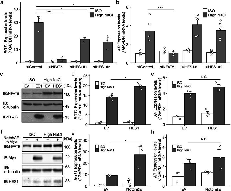
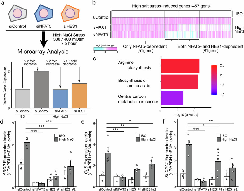
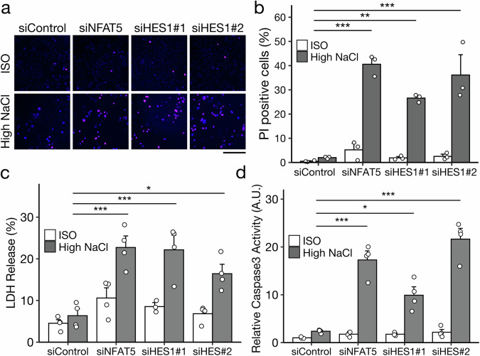
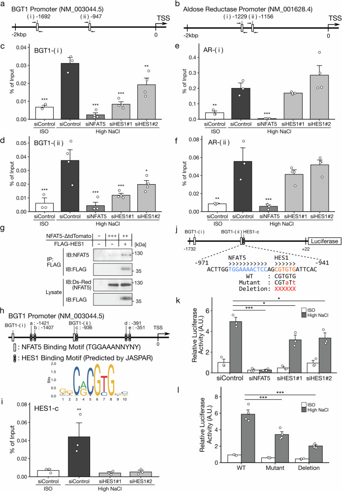
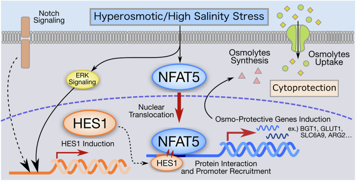
High salt conditions and subsequent hyperosmolarity are injurious cellular stresses that can activate immune signaling. Nuclear factor of activated T-cells 5 (NFAT5) is an essential transcription factor that induces osmoprotective genes such as aldose reductase (AR) and betaine-GABA transporter 1 (BGT1). High salt stress-mediated NFAT5 activation is also reported to accelerate the inflammatory response and autoimmune diseases. However, the systemic regulation of NFAT5 remains unclear. Here, we performed a genome-wide siRNA screen to comprehensively identify the regulators of NFAT5. We monitored NFAT5 nuclear translocation and identified one of the Notch signaling effectors, Hairy and enhancer of split-1 (HES1), as a positive regulator of NFAT5. HES1 was induced by high salinity via ERK signaling and facilitated NFAT5 recruitment to its target promoter region, resulting in the proper induction of osmoprotective genes and cytoprotection under high salt stress. These findings suggest that, though HES1 is well known as a transcriptional repressor, it positively regulates NFAT5-dependent transcription in the context of a high salinity/hyperosmotic response.


Conflict of interest statement

The authors declare no competing interests.

Figures

Fig. 1
Fig. 1. HES1 promotes NFAT5 nuclear translocation under high salt stress.
a, b A genome-wide siRNA screen to search for regulators of NFAT5 nuclear translocation under high salinity/hyperosmotic stress, 500 mOsm; 3 h. The dash line presented the value of NFAT5 nuclear translocation in non-target control siRNA-treated cells. b Notch-related genes in the positive hits whose knockdown attenuated NFAT5 nuclear translocation more than c-Abl1 knockdown. The vertical line shows the NFAT5 TL ratio, and the horizontal line shows the rank of genes. c KEGG pathway analysis of the 1291 positive hits. d, e Effect of HES1 depletion on NFAT5 nuclear translocation. Representative images of NFAT5-ΔtdTomato in the HeLa cells treated with siRNAs under ISO and high NaCl, 500 mOsm; 3 h. Blue and green lines represent the borders of the nuclear and whole-cell regions, respectively. The scale bar represents 10 µm. NT Non-transfected control. e NFAT5 TL ratio calculated from the fluorescent images (n = 3, analyzing 400–800 cells in each experiment). f, g Effect of HES1 depletion on the subcellular distribution of endogenous NFAT5 in HeLa cells under ISO and high NaCl, 400 mOsm; 3 h. f LAMIN A/C and β-tubulin are nuclear and cytoplasmic marker proteins, respectively. NFAT5 TL ratio calculated from the band intensity (g, n = 3). In the bar graphs, individual values (white points) and the mean ± SEM are presented. *p < 0.05, ***p < 0.001. ISO, 300 mOsm; high NaCl, 500 mOsm in (d, e) and 400 mOsm in (f, g); 3 h. See also Supplementary Fig. 1.
Fig. 2
Fig. 2. HES1 is induced by high salt stress via ERK signaling.
a Time course of HES1 protein expression in HeLa cells under ISO and high NaCl conditions, 400 mOsm. The bar graph indicates HES1 protein amount at 6 h after high NaCl stimuli (n = 4). b mRNA expression of Hes1 in HeLa cells at 5 h after a high NaCl stimulus, 400 mOsm (n = 5). mRNA expression in each sample was normalized to the mean expression of samples under isoosmotic conditions. c, d Effect of the ERK signaling inhibitor U0126 on high salt stress-induced HES1 protein under ISO and high NaCl conditions, 400 mOsm. Cells were pretreated with 5 µM U0126 for 30 min. DMSO: dimethyl sulfoxide. The bar graph indicates HES1 protein amount at 6 h after osmotic stimuli (c, n = 3) and mRNA expression (d, n = 3). mRNA expression in each sample was normalized to the mean expression of samples with DMSO treatment under isoosmotic conditions. e, f Effect of exogenous HES1 expression on NFAT5 nuclear translocation at 3 h after ISO and high NaCl stimuli, 400 mOsm. Representative images of NFAT5Δ-tdTomato in HeLa cells under isoomolarity. The scale bar represents 10 µm. EV: empty vector, a negative transfection control (e). NFAT5 TL ratio calculated from the fluorescent images (f, n = 3–5, analyzing 40–80 cells in each experiment). In the bar graphs, individual values (white points) and the mean ± SEM are presented. *p < 0.05, **p < 0.01, ***p < 0.001. ISO, 300 mOsm; high NaCl, 400 mOsm. See also Supplementary Fig. 2.
Fig. 3
Fig. 3. HES1 regulates the induction of a NFAT5 target gene, BGT1, but not that of another one, Aldose Reductase under high salt stress.
a, b Effect of HES1 depletion on the NFAT5 target genes BGT1 (a, n = 3) and Aldose Reductase (AR) (b, n = 5) at 7.5 h after ISO and high NaCl stimuli, 400 mOsm. mRNA expression in each sample was normalized to the mean expression of samples with control siRNA under isoosmotic conditions. c Expression of HES1 and NFAT5 in the HeLa cells transiently transfected with the EV or FLAG-HES1 construct at 6 h after ISO and high NaCl stimuli, 400 mOsm. d, e Effect of exogenous HES1 expression on the NFAT5 target genes BGT1 (d, n = 4) and AR (e, n = 4). f Endogenous HES1 expression in the HeLa cells transfected with a constitutively active Notch1 (NotchΔE-6Myc) construct at 6 h after ISO and high NaCl stimuli, 400 mOsm. g, h Effect of exogenous NotchΔE expression on the NFAT5 target genes BGT1 (g, n = 3) and AR (h, n = 3) at 7.5 h after ISO and high NaCl stimuli, 400 mOsm. In the bar graphs, individual values (white points) and the mean ± SEM are presented. N.S., not significant; *p < 0.05, **p < 0.01, ***p < 0.001. ISO, 300 mOsm; high NaCl, 400 mOsm. See also Supplementary Fig. 3.
Fig. 4
Fig. 4. HES1 promotes the induction of multiple NFAT5 target genes under high salt stress.
a Schematic model showing the design of the microarray analysis and gene classification. mRNA expression in HeLa cells treated with siControl, siNFAT5 and siHES1were analyzed at 7.5 h after ISO and high NaCl stimuli, 400 mOsm. b Heatmap showing the log fold change in the mRNA expression of high salt stress-induced genes at 7.5 h after a high NaCl stimulus, 400 mOsm. The expression under high salt conditions with each siRNA was compared with that under isoosmotic condition with siControl. c KEGG pathway analysis of the both NFAT5- and HES1-dependent genes (81 genes) in the microarray analysis. Effect of HES1 depletion on the both NFAT5- and HES1-dependent genes identified by microarray analysis at 7.5 h after ISO and high NaCl stimuli, 400 mOsm; ARG2 (d, n = 4), GLS (e, n = 5) and SLC2A1 (f, n = 5). In the bar graphs, individual values (white points) and the mean ± SEM are presented. N.S. not significant; *p < 0.05, **p < 0.01, ***p < 0.001. ISO, 300 mOsm; high NaCl, 400 mOsm. See also Supplementary Fig. 3.
Fig. 5
Fig. 5. HES1 has cytoprotective functions against high salt stress.
ad Effect of HES1 depletion on high salt stress-induced cell death. a, b HeLa cells treated with siRNAs were analyzed with Propidium iodide, PI staining assay at 24 h after ISO and high NaCl stimuli, 400 mOsm (a, representative images). Percentage of PI positive cells was calculated with image-based quantification (b, n = 3, analyzing 5000-10,000 cells in each experiment). c HeLa cells treated with siRNAs were analyzed with lactate dehydrogenase (LDH) release assay at 15 h after ISO and high NaCl stimuli, 400 mOsm (n = 4). d HeLa cells treated with siRNAs were analyzed with caspase 3 activity assay at 9 h after ISO and high NaCl stimuli, 400 mOsm. Caspase 3 activity in each sample was normalized to the mean activity of samples with control siRNA under isoosmotic conditions (n = 4). In the bar graphs, individual values (white points) and the mean ± SEM are presented. N.S. not significant; *p < 0.05, **p < 0.01, ***p < 0.001. ISO, 300 mOsm; high NaCl, 400 mOsm. The scale bar represents 100 µm. See also Supplementary Fig. 3.
Fig. 6
Fig. 6. HES1 regulates BGT1 promoter activity through the recruitment of NFAT5 to the target motif.
Schematic models showing the promoter regions of the NFAT5 target genes BGT1 (a) and Aldose Reductase (AR) (b). Each promoter region has two NFAT5-binding motifs, (i) and (ii), presented as white boxes. cf ChIP analysis investigating the effect of HES1 depletion on the DNA binding of NFAT5 to each motif, BGT1-(i) and –(ii) and AR-(i) and –(ii) at 7.5 h after ISO and high NaCl stimuli, 400 mOsm (n = 4). g Coimmunoprecipitation assay between over expressed NFAT5Δ-tdTomato and FLAG-HES1 in HEK293A cells. h A schematic model showing the prediction of HES1 binding motifs in the BGT1 promoter using JASPAR. The predicted sites of HES1 binding are presented as white boxes with x-markers. (i) ChIP analysis evaluating HES1 recruitment to a predicted site (HES1-c site) in the BGT1 promoter under high salt conditions at 5.5 h after ISO and high NaCl stimuli, 400 mOsm. j A schematic model showing the structure of the BGT1 reporter and mutations of the HES1 binding site introduced into the reporter. k Effect of HES1 depletion on BGT1 reporter activity at 9 h after ISO and high NaCl stimuli, 400 mOsm (n = 3). l Effect of a double point mutant (Mutant) and deletion mutant (Deletion) in the HES1 binding site on the BGT1 reporter activity (n = 4). Mutant sequences shown in (j).In the bar graphs, individual values (white points) and the mean ± SEM are presented. *p < 0.05, **p < 0.01, ***p < 0.001. ISO, 300 mOsm; high NaCl, 400 mOsm. See also Supplementary Fig. 4.
Fig. 7
Fig. 7. Schematic diagram of HES1 function in hyperosmotic/high salt stress response as a positive regulator of NFAT5.
HES1 potentiates DNA binding and nuclear translocation of NFAT5 as a positive regulator. Mechanistically, HES1 is induced by high salinity/hyperosmotic stress via ERK signaling and facilitates NFAT5 recruitment to its target promoter region through protein binding to NFAT5 and DNA binding to adjacent site of NFAT5 binding sites. The regulation is essential for the proper induction of osmoprotective genes and cytoprotection under high salt stress. Hes1 induced through the canonical Notch signaling can concomitantly contribute to NFAT5 nuclear translocation when it is activated.

References

    1. Alfieri, R. R. & Petronini, P. G. Hyperosmotic stress response: comparison with other cellular stresses. Pflugers Arch. Eur. J. Physiol.454, 173–185 (2007). - PubMed
    1. Burg, M. B., Ferraris, J. D. & Dmitrieva, N. I. Cellular response to hyperosmotic stresses. Physiol. Rev.87, 1441–1474 (2007). - PubMed
    1. Miyakawa, H., Woo, S. K., Dahl, S. C., Handler, J. S. & Kwon, H. M. Tonicity-responsive enhancer binding protein, a Rel-like protein that stimulates transcription in response to hypertonicity. Proc. Natl. Acad. Sci. USA.96, 2538–2542 (1999). - PMC - PubMed
    1. Cheung, C. Y. K. & Ko, B. C. B. NFAT5 in cellular adaptation to hypertonic stress – regulations and functional significance. J. Mol. Signal.8, 5 (2013). - PMC - PubMed
    1. Choi, S. Y., Lee-Kwon, W. & Kwon, H. M. The evolving role of TonEBP as an immunometabolic stress protein. Nat. Rev. Nephrol.16, 352–364 (2020). - PubMed