. 2024 Nov;635(8037):237-242.
doi: 10.1038/s41586-024-08015-6. Epub 2024 Oct 9.

Structural basis of mRNA decay by the human exosome-ribosome supercomplex

Affiliations

Structural basis of mRNA decay by the human exosome-ribosome supercomplex

Alexander Kögel et al. Nature. 2024 Nov.

Abstract

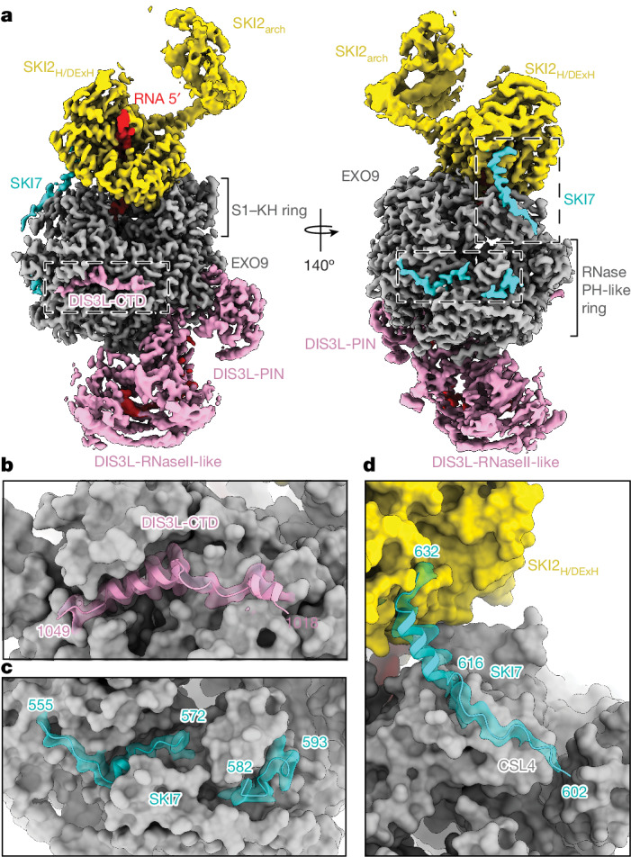
The interplay between translation and mRNA decay is widespread in human cells1-3. In quality-control pathways, exonucleolytic degradation of mRNA associated with translating ribosomes is mediated largely by the cytoplasmic exosome4-9, which includes the exoribonuclease complex EXO10 and the helicase complex SKI238 (refs. 10-16). The helicase can extract mRNA from the ribosome and is expected to transfer it to the exoribonuclease core through a bridging factor, HBS1L3 (also known as SKI7), but the mechanisms of this molecular handover remain unclear7,17,18. Here we reveal how human EXO10 is recruited by HBS1L3 (SKI7) to an active ribosome-bound SKI238 complex. We show that rather than a sequential handover, a direct physical coupling mechanism takes place, which culminates in the formation of a cytoplasmic exosome-ribosome supercomplex. Capturing the structure during active decay reveals a continuous path in which an RNA substrate threads from the 80S ribosome through the SKI2 helicase into the exoribonuclease active site of the cytoplasmic exosome complex. The SKI3 subunit of the complex directly binds to HBS1L3 (SKI7) and also engages a surface of the 40S subunit, establishing a recognition platform in collided disomes. Exosome and ribosome thus work together as a single structural and functional unit in co-translational mRNA decay, coordinating their activities in a transient supercomplex.


Conflict of interest statement

The authors declare no competing interests.

Figures

Fig. 1
Fig. 1. Structure of the human cytoplasmic exosome–ribosome supercomplex.
a, Schematic representation of the organization of the human SKI7 domain, with the binding domains for SKI238 (SKI), EXO10 (EXO) and 40S identified in this study (see also b and Extended Data Fig. 1). b, Identification of the SKI-EXO-binding region of SKI7 by pull-down assays with purified recombinant SKI238 and EXO10 complexes through GFP-tagged SKI7 (fragments or full-length (FL), as indicated). Mixed recombinant proteins (input, top panel, lanes 1–5) and proteins retained on GFP beads (precipitate, bottom panel, lanes 6–10) were analysed by SDS–PAGE. c, Single-particle cryo-EM composite map showing the 80S ribosome with the 60S coloured in light blue, 40S in sand, SKI2 helicase in yellow, EXO9 subunits in grey, DIS3L in pink, SKI7 in cyan and RNA substrate in red. The overall resolution estimates of the focused reconstruction are 3.2 Å, 3.3 Å and 3.7 Å for the 80S, 40S and cytoplasmic helicase–exosome assemblies, respectively. d, Model of the cytoplasmic EXO10-SKI-80S complex shown in cartoon representation, coloured as in c. Density for RNA is shown as an omit map displayed in a 30-Å radius around the IRES in red.
Fig. 2
Fig. 2. General and distinctive features of the ribosome-bound cytoplasmic exosome.
a, Focused cryo-EM reconstruction of EXO10-SKI bound to the 40S subunit of the cytoplasmic EXO10-SKI-80S complex. Colours are as in Fig. 1c,d. Different domains of DIS3L and the EXO9 barrel are indicated. bd, Magnified views of specific architectural features discussed in the text. The structural models of the DIS3LCTD (b) and SKI7EXO (c,d) are shown in cartoon representation, fitted in the transparent cryo-EM density. The other parts of the complex are shown with the structural model in non-transparent surface representation.
Fig. 3
Fig. 3. The SKI2N38 gatekeeping module is an interaction platform.
a, Single-particle cryo-EM reconstruction of SKI2Δarch38 bound to SKI7SKI at a global resolution estimated at 3.4 Å. The SKI238 complex is in an open conformation, with ordered density only for the SKI2N38 gatekeeping module (SKI3 is in blue, SKI2N in yellow, SKI8IN and SKI8OUT in light and dark green, respectively, and SKI7 in cyan). b, Magnified view of the SKI7SKI-binding site on SKI3. Density is in transparent representation. Residues L454 and I458 mutated in the assay in Extended Data Fig. 4h are highlighted. The SKI3 TPR helices 29B, 30A and 30B correspond to residues 1105–1115, 1123–1134 and 1139–1150, respectively. c, Single-particle cryo-EM composite map coloured as in a and in Fig. 1c. The overall resolution estimates of the focused reconstruction are 3.4 Å, 4.1 Å and 6.5 Å for the 40S subunit, the SKI2N38 gatekeeping module and the cytoplasmic helicase–exosome assemblies, respectively. d, Magnified view of the interaction site between the SKI2N38 gatekeeping module (in cartoon representation) and the 40S subunit. For clarity, the 40S-interacting regions are shown in cartoon representation and all other 40S components are highlighted. Colours are as in c. Residues that are mutated in patients with THES (L1485, R1503 and L1505) are shown as purple spheres.
Fig. 4
Fig. 4. The cytoplasmic exosome–ribosome supercomplex is compatible with disome engagement.
a, Superposition of the EXO10-SKI-40S composite map (shown in Fig. 3c) with the structure of a mammalian disome stalled on an XBP1 reporter mRNA. The disome is shown in cartoon representation and coloured in light grey (Protein Data Bank (PDB): 7QVP). All other colours are as in Fig. 3c. b, Sucrose gradient profiles (10–50% sucrose gradient) (left) and anti-Flag western blots (right) of the disome-containing gradient fractions of Xbp1-XTEN stalled RNCs with the SKI2N38 gatekeeping module (blue), Xbp1-XTEN stalled RNCs with SKI2Δwedge38 (red) and stalled RNCs only (control; black). a.u., arbitrary units; NC, nascent chain. c, Schematic depiction. Left, closed state of the SKI238 complex. Right, structural analysis of the cytoplasmic exosome–ribosome supercomplex with the SKI238 complex in an activated open state. The mRNA substrate (red) is being channelled from the 80S through the SKI2 helicase to the exosome DIS3L ribonuclease for degradation. In the model, the architecture of the exosome–ribosome supercomplex in RNA-degradation mode is compatible with the geometry of a collided ribosome (in grey), with the SKI2N38 gatekeeping module wedging in the composite surface formed by the stalled and collided 40S subunits (also see Extended Data Fig. 8). The schematic was created with BioRender.com. Source Data
Extended Data Fig. 1
Extended Data Fig. 1. Biochemical characterization of the human cytoplasmic exosome–ribosome supercomplex.
a, Domain organization of human HBS1L and SKI7 with used constructs labelled according to the binding of 40S, SKI and EXO described here. b, Distribution of iBAQ values for indicated protein isoforms. N = 25 data points collated from 5 cell lines each treated separately by 5 proteases. Quartiles are represented by boxes with whiskers extending to the maxima or minima value within 1.5 times the interquartile range. Median values are shown as thick horizontal bars inside the boxes. Values falling further than 1.5 times the interquartile distance are considered outliers and are shown as dots. iBAQ values are plotted in log scale. c,d, GFP protein co-precipitation assays. GFP-tagged SKI7 fragments were mixed with SKI238 complex (c) and 40S or 60S subunits (d, left and right panels respectively). The Coomassie-stained 4–12% SDS- PAGE gel shows the input on the top and the pulled down protein precipitates at the bottom. e, RNA substrate design to trap an active cytoplasmic exosome–ribosome supercomplex. Ribosome structure (grey) bound to CrPV-IRES (red/orange) is from an inactive SKI238-80S. The RNA length from the P site to the ribosomal surface is ~10 nucleotides and that spanning EXO10-SKI ~ 50-55 nucleotides,. CrPV-IRES will be long enough to reach the DIS3L exoribonuclease if the 3′ pseudoknot is unwound (PK-1, ~33 nucleotides, orange). f, RNA-degradation experiments with CrPV-IRES to trap a cytoplasmic ribosome-exosome supercomplex. The left-hand panel shows the RNA-degradation time courses in a 12% UREA- PAGE imaged for the 5′ fluorogenic Broccoli aptamer. The middle panel shows their densitometric quantitation (technical replicates n = 3). Per time point all three individual measurement points are shown. Trend lines connect the respective median. Coomassie-stained 4–12% SDS- PAGE gel with the purified input proteins is on the right.
Extended Data Fig. 2
Extended Data Fig. 2. Cryo-EM data analysis of the human cytoplasmic exosome–ribosome supercomplex.
a, A Coomassie-stained 4–12% SDS–PAGE of the complexes used for the reconstitution of the cryo-EM sample. M, molecular weight marker. b, A representative cryo-EM micrograph of the human cytoplasmic exosome–ribosome supercomplex sample in A is shown. This image was recorded at 300 kV with a pixel size of 0.85 Å/pixel using a post-GIF K3 direct detector. c, 2D class averages of the exosome–ribosome supercomplex. d, Processing scheme of the single-particle cryo-EM dataset of the exosome–ribosome supercomplex sample resulting in focused 3D reconstructions for the EXO10-SKI-80S (80S subtracted)/map1 and the EXO10-SKI-80S (60S subtracted)/map2 as well as the full EXO10-SKI-80S (full map)/map3. These three maps were used to calculate the composite map shown in Fig. 1b. The EXO10-SKI-80S map low-pass filtered at 20 Å is shown in Extended Data Fig. 6b. Masks used for the subtraction of partial particle signal are shown in red. e, The FSC of the masked and unmasked independent half maps for the EXO10-SKI/map1 reconstruction were calculated in the RELION 3.1 post-processing routine and the map vs model FSC using phenix.mtriage. The FSC cut-off criteria of 0.5 and 0.143 are indicated by dotted lines. f, Angular sampling distributions of the EXO10-SKI/map1 reconstruction. Sampling angle data were plotted in 3° by 3° bins and sampling bins coloured according to particle number with yellow indicating more particles and blue fewer particles. g, Local resolution estimate of the EXO10-SKI/map1 reconstruction. Position of SKI2, EXO9 and DIS3L are indicated on the right. h, Close up on the RNA path within the EXO10-SKI-80S assembly. Model orientation and colours are analogous to Fig. 1c. The model is shown in transparent cartoon representation except for the RNA. The density around the RNA (≤3 Å distance) is shown in black mesh.
Extended Data Fig. 3
Extended Data Fig. 3. Distinctive structural features of the human cytoplasmic exosome.
a, Cryo-EM reconstruction focused on human EXO10-SKI/map1. Colours are as in Fig. 2a except the colouring of specific EXO9 subunits discussed in the text: RRP45 in light green, RRP46 in light blue, RRP45 in light orange and CSL4 in salmon. be, Magnified views of specific architectural features discussed in the text including DIS3LCTD (b) SKI7 residues 555 to 593 (c), RRP45CTD (d) and SKI7 residues 602 to 632 (e). The structural models of RRP45CTD, DIS3LCTD and SKI7EXO are fitted in the cryo-EM density, shown in a transparent representation. The other parts of the complex are shown with the structural model in surface representation (non-transparent).
Extended Data Fig. 4
Extended Data Fig. 4. Cryo-EM data analysis of the human SKI7–SKI238 complex.
a, Peak fraction of a Superose 6 gel filtration run (labelled by *; chromatogram on left) of the human SKI7-SKI2Δarch38 complex analysed on a Coomassie-stained 4–12% SDS–PAGE and later used to prepare SPA (single particle analysis) cryo-EM specimen. M, molecular weight marker. b, A representative cryo-EM micrograph of the human SKI7-SKI2Δarch38 sample in A. This image was recorded at 300 kV with a pixel size of 0.85 Å/pixel using a post-GIF K3 direct detector. c, 2D class averages of the human SKI7-SKI2Δarch38 complex. d, Processing scheme of the single-particle cryo-EM dataset of the human SKI7-SKI2Δarch38 sample resulting in a 3D reconstruction of the complex at an overall estimated resolution of 3.4 Å (“SKI7-SKI238”/map4). This map is shown in detail in Fig. 3a. e, Local resolution estimate of the SKI7-SKI238 reconstruction. Red coloration indicates areas where the local resolution is estimated to be highest. f, The FSC of the masked and unmasked independent half maps for the SKI7-SKI238 reconstruction were calculated in the RELION 3.1 post-processing routine and the map vs model FSC using phenix.mtriage. The FSC cut-off criteria of 0.5 and 0.143 are indicated by dotted lines. g, Angular sampling distributions of the SKI7-SKI238 reconstruction. The data were plotted in 3° by 3° bins and these sampling bins coloured according to particle number with yellow indicating more particles and blue fewer particles. h, Pull-down assays with purified recombinant SKI2N38 gatekeeping module via GFP-tagged SKI7SKI, either wild-type or with structure-based mutations.
Extended Data Fig. 5
Extended Data Fig. 5. Comparison of yeast and human SKI7.
a, Alphafold2 prediction of the human SKI238 complex bound to SKI7 (residues 430–465). Model is shown in two different orientations related by a 90° rotation around a vertical axis. Colours are analogous to Fig. 3a. The model is shown in cartoon representation (left) and surface representation (right). The two helices of SKI7 are labelled SKI7-H1 and SKI7-H2. Helix H2 corresponds to the well-defined helix in the map, is displayed in Fig. 3b and is the major binding determinant. b, Model of the ySki238 complex bound to ySki7 (PDB:8QCB). Colours of the individual subunits and orientation of the model are analogous to a. ySki7 is coloured in orange and the three helices are labelled ySki7-H1, ySki7-H2 and ySki7-H3. c, Multiple sequence alignments of SKI3 from different organisms show a lack of conservation at SKI7/ySki7 binding sites. (Organisms with Uniprot identifiers used for alignments: S. cerevisiae: P17883, S. arboricola: J8PXS5, S. pombe: O94474, D. melanogaster: Q6NNB2, D. rerio: A0A8M1RLS7, X. laevis: Q6DFB8, T. rubripes: H2RXP3, M. musculus: F8VPK0, H. sapiens: Q6PGP7). Conservation of residues is indicated by colour from white (variable) to purple (conserved). Binding sites of SKI7 and ySki7 helices are indicated above the alignments with cylinders. d, Multiple sequence alignments of the conserved C-terminal domain of SKI3 present in metazoans. (Organisms and Uniprot identifiers analogous to c excluding yeast species and D. melanogaster.).
Extended Data Fig. 6
Extended Data Fig. 6. Cryo-EM data analysis of the human EXO10-SKI-40S assembly.
a, Representative 2D class averages of the human EXO10-SKI-40S assembly. These 2D class averages stem from the EXO10-SKI-80S supercomplex data recorded at 300 kV with a pixel size of 0.85 Å/pixel using a post-GIF K3 direct detector. b, Left panel: Low-pass-filtered full reconstruction of the EXO10-SKI-80S /map3 (cut-off 20Å) displays a density feature previously unaccounted for (black box). Right panel: indicated map area after focused 3D classification on the unaccounted density feature followed by 3D refinement (low-pass filtered to 20 Å). The structure of the EXO10-SKI-40S assembly is placed by rigid-body fitting. Colours as in Fig. 1b with SKI7 in grey. Asterisks indicate unmodelled rRNA features. c, Processing scheme of the EXO10-SKI-40S single-particle cryo-EM data resulting in 3D reconstructions of the EXO10-SKI-40S (full map)/map6 (at an estimated resolution of 3.4 Å) and focused reconstructions of the human SKI2N38 gatekeeping module (map5; at an estimated resolution of 4.1 Å) and the EXO10-SKI assembly (map7; at an estimated resolution of 6.5 Å). These reconstructions were used to calculate a EXO10-SKI-40S composite map shown in detail in Fig. 3. Map8 is a control for the presence of SKI2N38 gatekeeping module in the particles containing well-ordered EXO10-SKI. Masks used for the subtraction of partial particle signal are shown in blue.
Extended Data Fig. 7
Extended Data Fig. 7. Quality indicators of the SPA cryo-EM reconstructions of the human EXO10-SKI-40S assembly.
a, The FSC of the masked and unmasked independent half maps for the human 40S-bound SKI2N38 gatekeeping module reconstruction (map5) were calculated in the RELION 3.1 post-processing routine and the map vs model FSC using phenix.mtriage. The FSC cut-off criteria of 0.5 and 0.143 are indicated by dotted lines. b, Angular sampling distributions of the human 40S-bound SKI2N38 gatekeeping module reconstruction (map5). Sampling angle data were plotted in 3° by 3° bins and sampling bins coloured according to particle number with yellow indicating more and blue fewer particles. c, Local resolution estimate of the reconstruction focused on the 40S-bound SKI2N38 gatekeeping module (map5) with red indicating areas where the local resolution is estimated to be highest. Selected areas are labelled. d, The FSC of the masked and unmasked independent half maps for the human 40S-bound EXO10-SKI reconstruction (map7) were calculated in the RELION 3.1 post-processing routine and the map vs model FSC using phenix.mtriage. The FSC cut-off criteria of 0.5 and 0.143 are indicated by dotted lines. e, Angular sampling distributions of the human 40S-bound EXO10-SKI reconstruction (map7). Sampling angle data were plotted in 3° by 3° bins and sampling bins coloured according to particle number with yellow indicating more and blue fewer particles. f, Local resolution estimate of the reconstruction focused on the 40S-bound EXO10-SKI assembly (map7) with red indicating areas where the local resolution is estimated to be highest. Selected areas are labelled. g, Low-pass-filtered control EXO10-SKI-40S reconstruction (map8; cut-off 20Å) in two orientations displays well-ordered density for 40S, SKI2N38 gatekeeping module and EXO10-SKI. Colours as in Fig. 3c.
Extended Data Fig. 8
Extended Data Fig. 8. Structural features of the interactions between SKI7, SKI238, the exosome and the ribosome.
a, Structural superposition of the human EXO10-SKI-40S assembly on a stalled disome (PDB:7QVP). Stalled 80S ribosome and EXO10-SKI coloured similar to Fig. 1b, collided ribosome in grey, SKI2N38 gatekeeping module analogues to Fig. 3a and the RNA in red. The disome unit is shown in surface representation, SKI238 and the cytosolic exosome in cartoon representation. b, Detailed view of the potential contact sites between human SKI2N38 gatekeeping module and the 40S subunits of the stalled (grey) and the collided (sand) ribosome. Based on the superposition, the ribosomal proteins eS31 (blue), eS12(orange) and helix H33 of the 18S rRNA of the collided ribosome were identified as potential interactions sites for SKI8IN. In contrast, SKI2H interacts with its RecA2 with uS3, uS12, eS10, and 18S rRNA helix 16 and with its SKI2arch with uS3, uS10, and 18S rRNA helix 41. c, Detailed view of the superposition of the SKI2N38 gatekeeping module and the SKI2H helicase module on the collided disome structure (PDB:7QVP), focusing on known ubiquitination sites on the 40S subunits. Purple spheres indicated the approximate ubiquitination sites of the ribosomal proteins uS3, eS10 and uS10. For eS10 and uS10, the ubiquitination sites are in structurally flexible regions and not modelled. Therefore, the closest proximal residues are highlighted as spheres.
Extended Data Fig. 9
Extended Data Fig. 9. Interactions between collided disomes and the SKI complex.
a, Schematic of the in vitro translation and isolation protocol of ribosome nascent chain complex (RNC) using the Xbp1-XTEN mRNA in rabbit reticulocyte lysate (RRL). The cartoon on the right illustrates the stalling/colliding of the RNCs on the Xbp1-XTEN mRNA. Note that the RNCs contain both a 3xFLAG tag (used in Western blots here) and an 8xHis tag (used for purification purposes here) on the nascent chain. b, 10–50% sucrose gradient profiles (panel on the left) and anti-FLAG Western blots (panel on the right) of the gradient fractions of stalled RNCs only (b1), Xbp1-XTEN stalled RNCs with SKI2Δwedge38 (b2), Xbp1-XTEN stalled RNCs with SKI2N38 gatekeeping module (b3) and SKI2N38 gatekeeping module only (b4). Flag positive control is SKI2N38 gatekeeping module in panels b1, b3 and b4 as well as SKI2Δwedge38 in b2. Both SKI3 and the nascent chain are Flag-tagged. Source Data

References

    1. Wu, Q. & Bazzini, A. A. Translation and mRNA stability control. Annu. Rev. Biochem.92, 227–245 (2023). - PubMed
    1. Morris, C., Cluet, D. & Ricci, E. P. Ribosome dynamics and mRNA turnover, a complex relationship under constant cellular scrutiny. Wiley Interdiscip. Rev. RNA12, e1658 (2021). - PMC - PubMed
    1. D’Orazio, K. N. & Green, R. Ribosome states signal RNA quality control. Mol. Cell81, 1372–1383 (2021). - PMC - PubMed
    1. Hu, W., Sweet, T. J., Chamnongpol, S., Baker, K. E. & Coller, J. Co-translational mRNA decay in Saccharomyces cerevisiae. Nature461, 225–229 (2009). - PMC - PubMed
    1. Pelechano, V., Wei, W. & Steinmetz, L. M. Widespread co-translational RNA decay reveals ribosome dynamics. Cell161, 1400–1412 (2015). - PMC - PubMed

MeSH terms

LinkOut - more resources